Age-related macular degeneration (AMD) affects the macula, the centre of the retina, and with it central vision. Dry AMD is common and changes slowly; wet AMD can change vision quickly and is treated with eye injections.
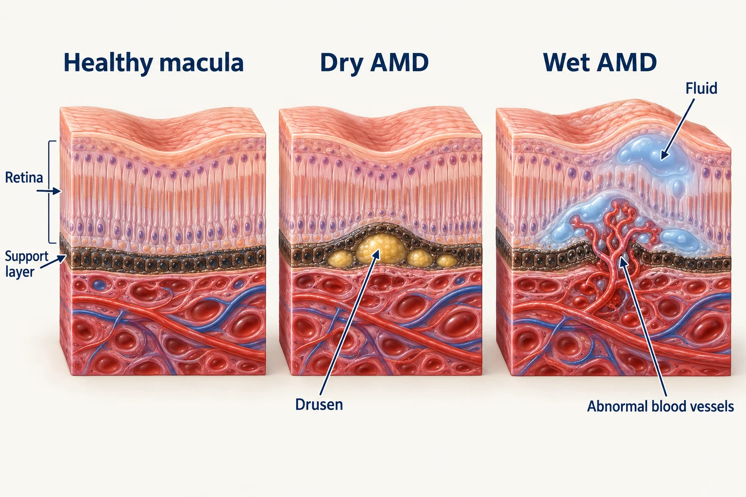
Changes beneath the macula. Simplified cross-sections compare healthy tissue, dry AMD and wet AMD. Actual changes vary between eyes.
The macula is the small central area of the retina that gives sharp, detailed vision for reading, driving and recognising faces.
Age-related macular degeneration (AMD) is a condition in which this central area wears and changes with age. It affects central vision, not the edges, so people rarely lose their sight completely, but reading, faces and fine detail can become difficult.
There are two forms. Dry AMD is the more common and usually progresses slowly, as deposits called drusen build up under the macula and the retinal cells gradually thin. Wet AMD is less common but can change vision quickly, when fragile new blood vessels grow under the macula and leak fluid or blood. Dry AMD can turn into wet AMD, which is why any new distortion needs a prompt check.
Affects central vision, not side vision
Dry AMD: common, slow, monitored
Wet AMD: less common, faster, treated with eye injections
New distortion or a central smudge needs prompt review
Smoking is the main avoidable risk factor
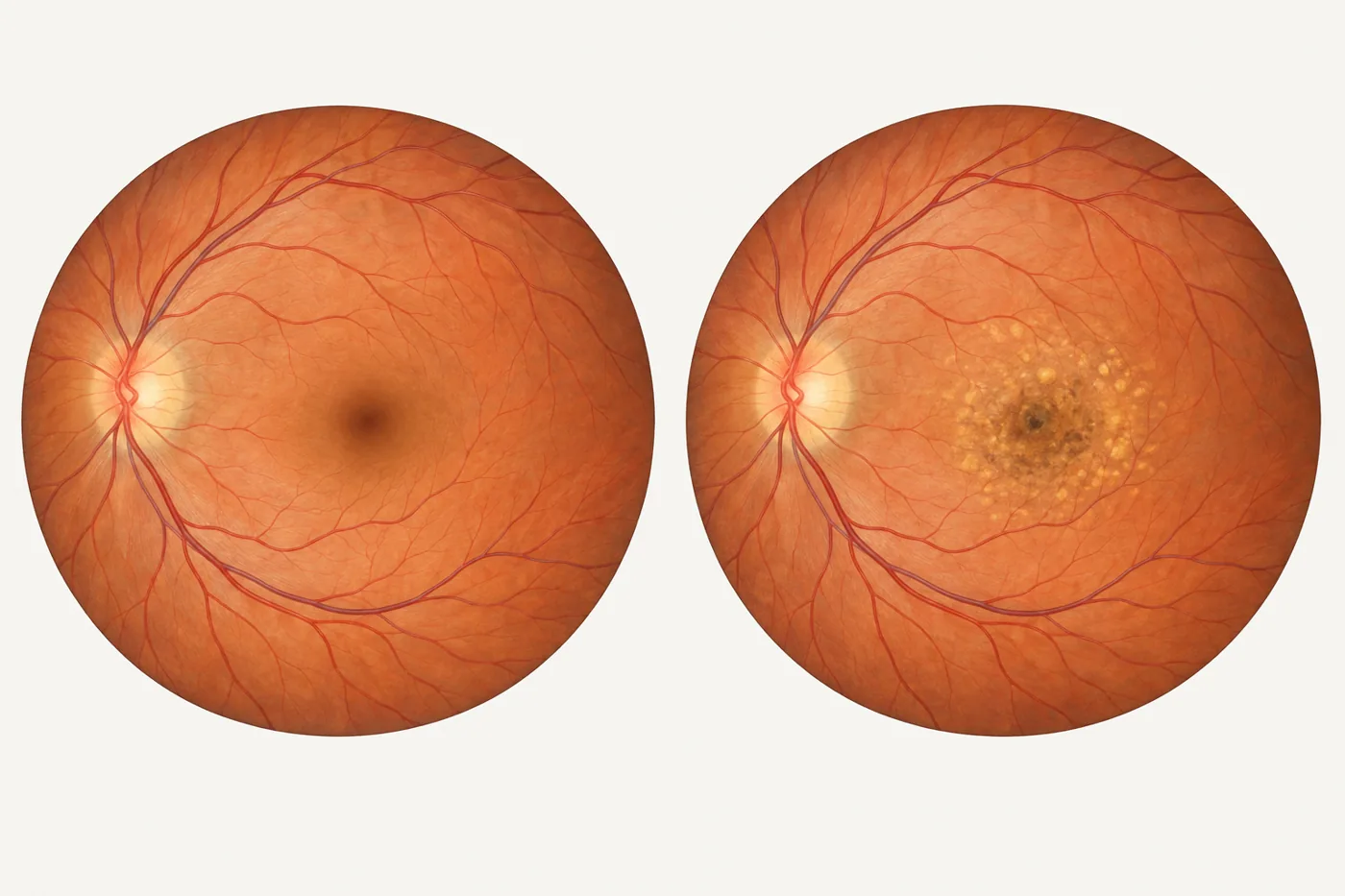
Left: a healthy macula. Right: yellow deposits (drusen) and pigment change in macular degeneration.
What are the symptoms of macular degeneration?
Because the macula is at the centre of the retina, the symptoms sit in the middle of whatever you look at.
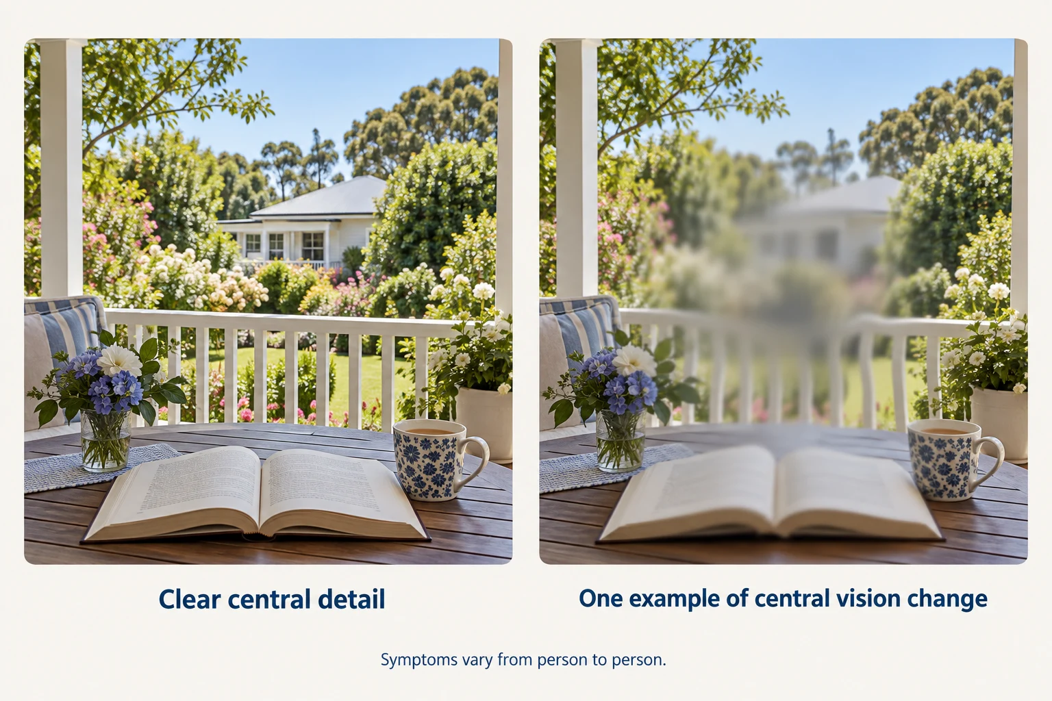
How central vision may be affected. This is one illustration of central blur and distortion, not a prediction of an individual person’s vision.
Lines of print, door frames, tiles or the edge of a road look wavy, bent or kinked, most often in one eye.
⇔
ClearWith AMD
A central smudge
A blurred, grey or missing patch in the centre of vision, so the middle of a face or a word is hard to make out while the edges stay clear.
⇔
ClearWith AMD
Fading detail and colour
Print needs more light or magnification, colours look less vivid, and faces can be harder to recognise across a room.
A new central blur, distortion or missing area needs prompt assessment. It may indicate wet AMD or another eye problem. Contact the clinic when you notice a change rather than waiting for your next appointment.
Who is at risk of macular degeneration?
Age
Most common from the sixties onward and increasingly so with each decade.
Smoking
The strongest avoidable risk factor. Stopping helps at any age.
Family history
A parent or sibling with AMD raises the likelihood, so relatives are encouraged to have their macula checked.
Blood pressure and heart health
Cardiovascular risk factors are linked with AMD, and looking after them helps the eye too.
Diet and weight
A diet rich in green leafy vegetables and fish is associated with lower risk. Supplements are discussed individually.
AMD in the other eye
Once one eye is affected, the other is watched more closely.
How is macular degeneration assessed?
Most of the assessment is looking, measuring and scanning. It does not hurt.
After a vision test, dilating drops enlarge the pupil so the macula can be examined properly. An OCT scan (optical coherence tomography), which uses light to map the layers of the retina in fine detail, shows drusen, thinning, fluid or bleeding under the macula, and is repeated over time to track change. Retinal photographs are usually taken, and in some eyes a dye test (angiography) is used to show leaking vessels.
Dr Chen explains what the scan shows, which form of AMD is present, and what that means for your vision and treatment.
How can I check my vision at home?
An Amsler grid is a square of straight lines with a dot in the middle
Cover one eye, wear your reading glasses and look at the centre dot in good light
Notice any wavy, blurred or missing lines, then repeat with the other eye
Any new change: phone the clinic rather than wait for the next visit
A self-check between visits does not replace an examination.
What do eye injections for macular degeneration involve?
An intravitreal injection places medication directly into the vitreous, the clear gel that fills the eye, so it reaches the macula at a useful concentration.
For wet AMD the medications block the signal that drives abnormal blood vessels to grow and leak under the macula. They are not a one-off. Treatment is given as a course: usually a series of injections about a month apart at first, then at intervals set by how the macula looks on the OCT scan. Many people continue treatment, often at a longer interval, for years.
Dr Chen was principal investigator in international clinical trials in macular degeneration, including the VIEW2 trial. He discusses the medication, the schedule and the trade-offs with each patient before treatment begins.
A vision check and an OCT scan of the macula
Anaesthetic drops and an antiseptic wash of the eye surface
The injection itself takes seconds; most people feel pressure rather than pain
Grittiness, redness or a small bleed on the white of the eye for a day or two is common
The interval to the next injection is set at each visit
Limitations: the injections treat wet AMD but do not cure it; they do not help dry AMD (geographic atrophy treatment is described below); treatment continues for as long as the eye needs it; and, as with any injection into the eye, there is a small risk of infection, retinal detachment or raised pressure. Suitability is discussed at the consultation.
How are wet and dry macular degeneration treated?
01
Wet AMD: a course of eye injections
Wet AMD is treated with intravitreal injections, described above. The aim is to dry the leak under the macula and protect the vision you have; the interval between injections is set by the OCT scan at each visit. Suitability, the medication and the schedule are discussed individually.
02
Dry AMD: monitoring and lifestyle
No injection or operation reverses dry AMD. For some people with geographic atrophy, the advanced dry form, injections that slow its growth may be considered, as described below. Care centres on regular monitoring with OCT, not smoking, a diet rich in green leafy vegetables and fish, looking after general health and, for some people, a specific vitamin formulation where the examination suggests it may help. Above all, any new distortion or smudge needs prompt review, because it may signal a change to the wet form.
03
What helps with everyday vision?
Good lighting, magnification, larger print and strong contrast all help make the most of the vision you have. Low-vision services can be arranged when they are useful, and driving requirements are discussed openly.
Suitability for any treatment is decided at the consultation, based on the examination and scans.
Is there a treatment for geographic atrophy?
Geographic atrophy is the advanced form of dry AMD, in which patches of retinal cells under the macula are lost. For some people, eye injections can now slow how quickly those patches grow. They do not bring back vision that has already been lost, and they do not make vision feel sharper.
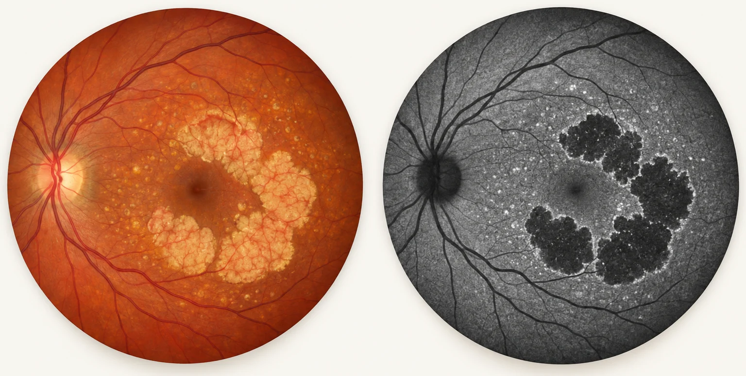
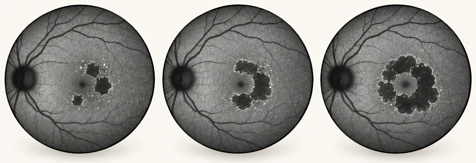
The patches are seen most clearly with autofluorescence imaging, which records the natural glow of the retina’s supporting cells: where those cells have gone, the scan is black. An OCT scan shows the same thinning in cross-section. Comparing today’s scans with earlier ones shows how quickly the patches are growing. Some inherited retinal conditions can look similar, so confirming that the atrophy is due to AMD comes first.
The same eye, two ways. Left, a colour photograph: the atrophy looks pale. Right, an autofluorescence scan: the same areas are black, with a brighter edge where they tend to spread.
Why can the eye chart look normal with geographic atrophy?
Geographic atrophy often starts beside the very centre of vision and grows around it before reaching it. The eye chart tests that tiny central point one letter at a time, so it can stay good for years.
Everyday tasks tell a different story: the middle of words breaks up when reading, faces take longer to recognise, subtitles are tiring and dim light becomes difficult. It is worth telling Dr Chen about these changes even when the chart reading is good, because they help show how much the atrophy is affecting daily life.
⇔
ClearWith geographic atrophy
Reading with geographic atrophy. The word being looked at can stay clear while parts of the words around it drop out.
How fast does geographic atrophy grow?
It varies a great deal from one eye to another. Some patches creep slowly over many years; others spread faster. Earlier scans are the best guide, because they show how a particular eye has behaved.
Growth tends to be faster when there are several separate patches, when the patches lie further from the centre or have irregular edges, when both eyes are affected, and when a type of deposit called reticular pseudodrusen is present. A single small patch usually grows more slowly. Nobody can give an exact timetable for an individual eye, but these features help judge the risk.
Growth over time. Left to right, autofluorescence scans of the same eye at visits a few years apart: separate patches enlarge and merge around the centre.
What can geographic atrophy treatment do?
Dr Chen often compares it with a bushfire. The black area is land that has already burned, and no treatment can regrow it. The injections aim to slow how fast the fire spreads into the land around it.
In the main clinical trials, the patches grew on average roughly 15 to 20 per cent more slowly than in untreated eyes, measured on scans. The trials did not show an improvement in eye chart vision. The benefit is real but modest, and it is usually seen on the scan rather than felt. That is very different from wet AMD, where injections often dry the retina and vision can improve.
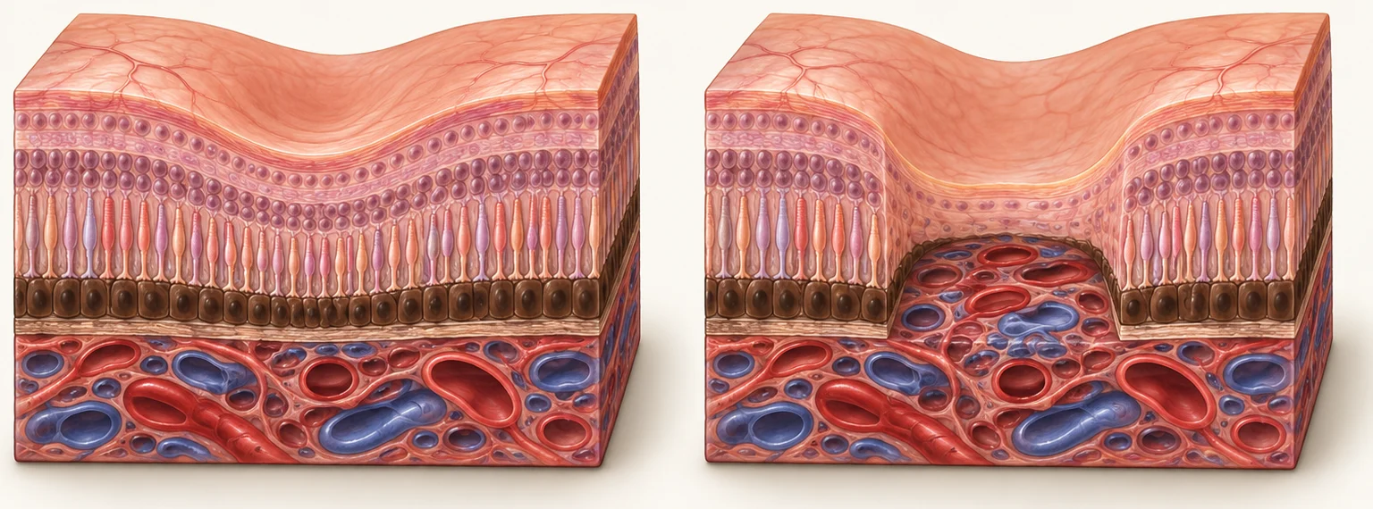
What is lost. Left, a healthy macula. Right, geographic atrophy: the light-sensing cells and their supporting layer are gone in a sharply edged zone. Treatment cannot restore that zone; it aims to slow its spread at the edge.
Who may be suitable for geographic atrophy injections?
The centre of vision (the fovea) is still working
The patches are close enough to threaten it
Enough central vision remains for slowing to matter in daily life
The aim, to slow loss rather than improve sight, is clearly understood
Treatment is less likely to help once the centre has already been lost, or when another condition, such as advanced glaucoma, is the main cause of poor vision. The other eye, getting to regular appointments and cost all form part of the decision.
What does geographic atrophy treatment involve?
An injection into the eye every month or every other month, depending on the medication
Ongoing treatment, for as long as it remains worthwhile
The same steps as other eye injections: anaesthetic drops, an antiseptic wash and an injection that takes seconds
Regular OCT scans, including to check for wet AMD
What if I decide not to have injections?
Choosing not to have injections is a reasonable, informed decision, and it is not the end of care. Scans continue, wet AMD is still watched for, and low-vision support with lighting, magnification, contrast and reading strategies helps from the start.
What are the risks of geographic atrophy injections?
Wet AMD. Treated eyes are more likely to develop wet AMD. In the main trials, roughly 7 to 12 in every 100 treated eyes developed it within one to two years, compared with about 3 to 4 in every 100 untreated eyes. Wet AMD needs its own injections, so regular scans continue.
Inflammation of the retinal blood vessels. Rare, in the order of 1 in 10,000 injections, but it can seriously damage vision. It has occurred most often after the first injection, usually within about two weeks.
The risks of any eye injection: infection inside the eye, inflammation, retinal detachment and raised eye pressure.
The burden of treatment: repeated visits, usually every one or two months, for an effect that is usually not felt.
Pain, increasing redness, new floaters or a sudden drop in vision after an injection need urgent review.
Is geographic atrophy treatment covered by the PBS?
No. Two medications are registered by the Therapeutic Goods Administration for geographic atrophy in Australia, but neither is subsidised on the Pharmaceutical Benefits Scheme. The cost of the medication is currently prohibitively expensive for most patients.
Geographic atrophy: managing patient expectations in the era of treatment
Written for optometrists, Dr Chen’s article explains what the new geographic atrophy treatments can and cannot do, which patients benefit from an early opinion, and why the goal of treatment needs to be clear before the first injection.
Continuing education article for eye-care professionals · mivision website
Can I have cataract surgery with macular degeneration?
Macular degeneration is one of the conditions that changes the cataract conversation. It can affect the lens choice, the timing and what to expect afterwards, so the plan takes both conditions into account. Dr Chen has written about cataract surgery for people with macular degeneration.
Published in Optometry Connection · May 2024
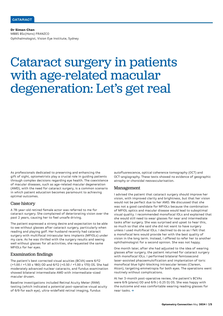
Cataract surgery in patients with age-related macular degeneration: Let’s get real
This article focuses on people who have both cataracts and age-related macular degeneration. Dr Chen explains why macular health, visual quality and realistic expectations guide the choice of lens for these patients.
Original published article for eye-care professionals · 3 pages · PDF, 106 KB
What should I ask Dr Chen about macular degeneration?
Which form of AMD do I have, and in which eye?
What does my OCT scan show today, and what will you compare it with next time?
If injections are suggested: which medication, how often at first, and how will the interval be decided?
What should I do between visits if I notice a change?
Are supplements appropriate for me at this stage?
How will AMD affect driving, reading and the things I do most?
Who do I phone if something changes suddenly?
Common questions about macular degeneration
Will I go blind?
AMD affects central vision. Side vision is preserved, so people keep the ability to get around, although reading and faces can become difficult when the centre is affected. Treatment for wet AMD aims to protect the vision you have.
How often are the injections?
Usually monthly at first, then the interval is set by how the eye responds on the OCT scan. Some eyes need injections for years; others can be spaced out. The schedule is reviewed at every visit.
Are the injections painful?
The eye is numbed with drops and the injection itself takes seconds. Most people describe pressure or a brief sting rather than pain, and some grittiness afterwards from the antiseptic.
Can dry AMD turn into wet AMD?
Yes, in a proportion of eyes, and usually with a noticeable change: new distortion or a central smudge. That is why self-checks between visits, and prompt review of any change, matter.
Should I take supplements?
Specific vitamin formulations have been studied in AMD and may be suggested for some people at a particular stage of the condition. They are not a treatment for everyone and are discussed individually.
Is macular degeneration hereditary?
Family history increases the risk, so close relatives are encouraged to have their macula checked at routine eye examinations.
Is there a treatment for geographic atrophy?
For some people, yes. Eye injections can slow how quickly the patches of geographic atrophy grow. They do not restore lost vision. Suitability depends on whether the centre of vision is still working and threatened by the patches.
Will geographic atrophy injections improve my vision?
No. They aim to slow further loss, not to improve sight. Many people notice no change in their vision even when treatment is working; the effect is measured on scans over time.
Where can I read more?
Longer articles by Dr Chen go into these questions in more detail, including what the research shows.
The difference between wet and dry AMD explains the medical names, how wet disease develops on underlying AMD, and which forms can be treated with injections.
How do I make an appointment with Dr Chen to discuss macular degeneration?
Appointments and enquiries are made through Vision Eye Institute Chatswood.
Vision Eye Institute confirms your appointment after you submit your request. The request form is not for emergencies. A referral is not strictly essential to request an appointment, but you need a valid one to claim a Medicare rebate for your consultation.