
Vitreous frills after vitrectomy: what are they and can they be treated?
A vitreous frill is a term used for the visible edge of gel remaining near the edge of the eye after vitrectomy. Some patients notice it as a moving peripheral strand, ripple or shadow after surgery for floaters. Observation may be appropriate once the retina has been checked; persistent, troublesome remnants can sometimes be treated with further surgery.

Published
What is a vitreous frill?
Vitrectomy removes vitreous, the clear gel behind the lens. It is not possible to remove 100% of the gel. A thin skirt of gel usually remains near the vitreous base, a ring of particularly firm attachment around the far edge of the retina, towards the front of the eye. In some eyes, the cut edge or strands of this remaining gel become visible.
“Frill” describes that residual edge. A peripheral shadow cannot be identified as a frill from its description alone.
The vitrectomy surgery guide explains the operation more generally. The important distinction here is between gel deliberately retained to protect delicate structures, a troublesome remnant that might be trimmed, and a different eye problem needing treatment.
New flashes, a sudden shower of floaters, a curtain or missing area of vision need urgent assessment. Do not assume they are a harmless frill.
What might a frill look like?
Patients describe a transparent ribbon, wavy edge, grey strand or flickering shadow that swings into peripheral vision when they move their eyes. Some notice it mainly against a bright sky, white screen or pale wall. Others find it distracting when reading, driving or changing their gaze between screens.
These descriptions are not a diagnostic checklist. A small visible remnant may be very intrusive even when the central vision chart result is good. Conversely, gel can remain without producing any noticeable symptom.
The article on how floaters cause symptoms explains why the position and movement of an opacity matter. The example below illustrates ordinary floater shadows; a peripheral frill can look quite different.
How ordinary floaters interrupt a bright view
Educational simulation of floater shadows, not a diagnostic picture of a frill or a prediction of surgery.
Why might younger patients notice this problem?
Younger eyes often have more formed gel, with attachments that have not loosened through the usual age-related separation process. Removing it can require more manipulation than removing gel that has already separated. Pulling on firmly attached vitreous can transmit traction to the retina and cause a tear.
There is also the natural lens to protect. The surgeon works through the space behind it, and contact with the lens can cause a cataract. Reaching the most anterior gel while keeping instruments safely away from the lens can be difficult.
These considerations may lead a surgeon to leave more peripheral or anterior gel, particularly in a younger eye. They provide a plausible explanation for frills being encountered in this group; they do not establish an age-specific probability. Published studies have not reliably measured how often symptomatic frills occur in younger versus older patients.
Age alone does not determine the operation. Existing vitreous separation, short-sightedness, lens status, retinal findings and the position of the troublesome opacities all matter.
Why not remove every last piece of vitreous?
The aim is to improve vision while protecting the retina and lens. The vitreous base is strongly attached, so trying to strip it completely is not the same as safely trimming gel projecting from it. Even an extensive vitrectomy generally leaves some tissue at these attachments.
You may encounter several terms when discussing surgery:
- Core or limited vitrectomy: removal focuses on troublesome gel, with some vitreous deliberately retained. The exact extent varies.
- PVD induction: the surgeon creates a posterior vitreous detachment, separating the back surface of the gel from the retina when it has not already separated.
- Peripheral shaving: the surgeon trims peripheral gel under direct visual control, sometimes with gentle pressure on the outside of the eye to improve the view.
These describe different parts of an operation. Inducing a PVD does not mean that every piece of anterior vitreous has been removed, and the word “full” does not guarantee freedom from residual symptoms. A limited approach and a more extensive approach involve different trade-offs. Ask what is planned for your particular eye rather than relying on a label.
Is a frill the same as floaters returning months later?
Not necessarily. Residual peripheral gel and a later posterior vitreous detachment are related but distinct possibilities.
After a limited vitrectomy without an existing complete PVD, retained posterior gel can separate later and produce new, more central floaters. A 2023 study documented this problem after limited vitrectomy. Its reported recurrence figures describe floaters associated with a new PVD; they are not rates of vitreous frills.
A symptom may also become noticeable only after early postoperative blur clears or when lighting and visual activities change. Timing alone cannot identify the cause. A new symptom several months after surgery still deserves assessment, particularly if it appears suddenly or includes flashes.
Will it settle without another operation?
It may become less noticeable, but this cannot be promised. Once a retinal problem has been excluded, allowing the eye to recover and reassessing the symptom can be sensible. Changes in the remnant's position, clearing of other postoperative changes and adaptation may each affect what a person notices.
There is no well-established timetable by which a true frill must disappear. Continuing to notice it early after surgery does not establish that it will be permanent.
Describe its impact in ordinary activities rather than repeatedly trying to provoke it against the brightest possible background. A short note about when it occurs, whether it has changed and what it stops you doing can make follow-up more useful. Do not wait for a routine appointment if warning symptoms develop.
How does the surgeon work out what is causing it?
Assessment starts with the history, a dilated examination of the vitreous and peripheral retina, and review of the original operation. It helps to know whether a PVD was present or induced, whether retinal tears were treated and whether air or gas was used.
Depending on the findings, ultrasound may help assess residual vitreous or a PVD, while an OCT scan examines the macula. A normal central OCT does not, by itself, explain or exclude a peripheral symptom.
Other possible explanations include ordinary residual floaters, postoperative inflammation or bleeding, a bubble edge, retinal traction or a retinal tear. After cataract surgery, lens-related arcs or shadows can also be considered. The article on negative dysphotopsia after cataract surgery describes one such symptom. Similar wording does not mean the underlying cause is the same.
Can surgery remove a troublesome frill?
Yes. A further vitrectomy can trim symptomatic residual vitreous in selected cases. The strongest reason to consider it is a persistent symptom that meaningfully affects daily life, with examination findings that offer a credible, treatable explanation.
The surgeon must decide what can be removed safely and whether other remaining vitreous needs attention. This can involve careful peripheral inspection and trimming; if posterior vitreous remains attached, the implications of separating it also need discussion. It is not a blind “clean-up” based on a patient's description alone.
Possible benefits must be balanced against another exposure to surgical risk. These include retinal tears or detachment, cataract progression or lens injury in an eye with its natural lens, bleeding, infection, pressure changes and macular swelling. Vision can be permanently damaged. A remnant can remain, symptoms may persist, and removing one visible edge does not guarantee perfectly clear vision.
What does vitrectomy for floaters involve?
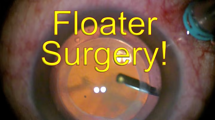
Dr Chen’s video shows real floater surgery. It illustrates vitrectomy, not a frill-specific revision or an expected outcome.
Watch on YouTube →Does previous cataract surgery make removal easier?
It can improve access. After cataract surgery, the natural lens has been replaced by a much thinner artificial lens. The surgeon no longer has to avoid damaging the natural lens and causing a cataract. This can make it easier to reach and trim anterior or peripheral vitreous.
The same advantage may apply when cataract surgery and vitrectomy are planned together for an eye that also has a cataract. The combined cataract and retinal surgery guide explains this approach.
The retinal attachments do not disappear, however. The lens implant, its supporting capsule and the retina still need protection. Previous cataract surgery therefore does not make frill removal risk-free or guarantee complete removal.
Removing a clear natural lens solely to gain access is a separate decision, particularly in a younger person who still uses it to focus up close. It should not be treated as an automatic step in managing a frill. For context, read why vitrectomy can cause cataracts.
What else do patients commonly ask?
Does a frill mean that my surgeon made a mistake?
No. Some vitreous is deliberately retained for safety, and a visible remnant does not by itself establish poor technique. There is no reliable comparative evidence showing that symptomatic frills are more common with less experienced floater surgeons. It is reasonable to seek an explanation of the original surgical choices or another retinal surgeon's opinion if symptoms remain troublesome.
Can a frill occur after a “full” vitrectomy?
Yes, peripheral or anterior remnants may remain even when a PVD has been induced and the main vitreous cavity cleared. “Full” is not a guarantee that every attachment has been removed. Discuss the actual operative findings and remaining gel rather than treating the term as a promise.
Can laser, eye drops or supplements remove it?
No eye drop or supplement has been established as a treatment that removes a postoperative vitreous frill. YAG laser used for selected floaters is not automatically suitable for gel near the lens or retina. Location, attachment and a clear view are critical, and good evidence specifically supporting laser treatment of postoperative frills is lacking.
Has the vitreous grown back?
Removed vitreous gel does not simply regenerate as the original gel. A later symptom may come from vitreous that remained, a change in its position or separation, or another postoperative cause. Examination is needed to distinguish these possibilities.
What should I ask before considering a second operation?
Ask what structure appears to explain your symptom; whether the retina is healthy; whether further observation is reasonable; what gel can be removed safely; how your lens status changes the plan; and what outcome would count as worthwhile improvement. Also ask about the possibility of persistent symptoms, complications and recovery restrictions.
Where can I read more?
When should I seek urgent advice?
Contact your surgeon urgently for new flashes, a sudden increase or shower of floaters, a dark curtain or missing area of vision, or a sudden reduction in sight. Increasing pain or redness after surgery also needs prompt advice.
Do not label a new shadow a frill and wait for it to settle. If your surgical team cannot be reached, seek urgent ophthalmic care or attend an emergency department. A previous vitrectomy does not eliminate the possibility of a retinal tear or detachment.
Sources
- Sebag J. Vitreous and vision degrading myodesopsia. Progress in Retinal and Eye Research. 2020;79:100847. Full review .
- Sebag J, Yee KMP, Nguyen JH, Nguyen-Cuu J. Long-term safety and efficacy of limited vitrectomy for vision degrading vitreopathy resulting from vitreous floaters. Ophthalmology Retina. 2018;2:881–887. Study .
- Boneva SK and colleagues. Recurrent floaters after limited vitrectomy for vision degrading myodesopsia. Retina. 2023;43:1114–1121. PubMed record .
- American Society of Retina Specialists. Vitrectomy for floaters .
- American Society of Retina Specialists. Retinal detachment .
- University of Utah, Moran CORE. Vitreous anatomy and pathology .
- University of Michigan, The Eyes Have It. Eye in cross section .