
What is the Yamane technique for fixing a lens implant?
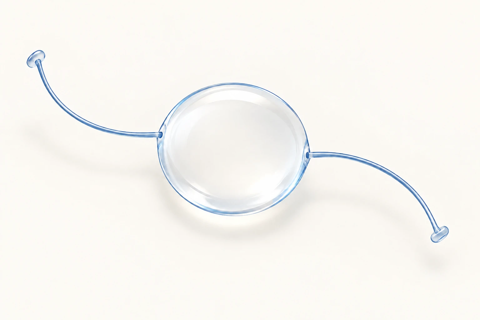
The Yamane technique secures an artificial lens inside the eye when the eye’s usual lens support is missing or too weak. The lens sits behind the coloured iris. Its two fine supporting arms are anchored in the white wall of the eye, without permanent stitches holding the implant in place.

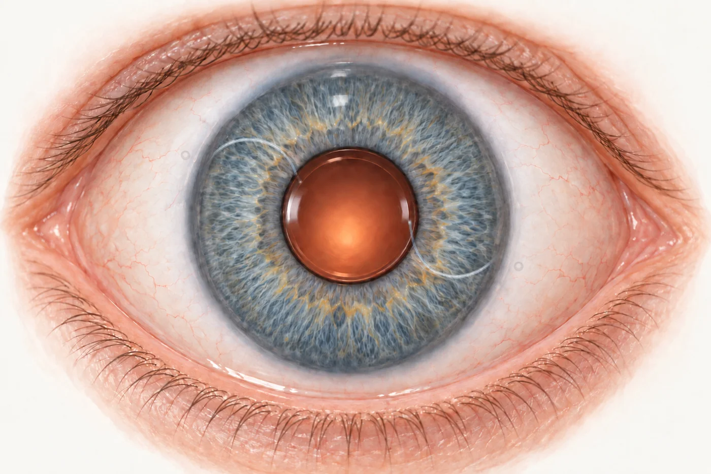
It can be useful after a complicated cataract operation, an eye injury or movement of an older implant. However, it is one of several ways to support a lens. The most suitable method depends on the condition of the eye and the implant.
The central disc focuses light. The two supporting arms are called haptics. Their enlarged tips, or flanges, help anchor the implant.
Why might a lens implant need extra support?
During ordinary cataract surgery, the artificial lens usually sits inside the thin capsule that previously surrounded the natural lens. Fine fibres, called zonules, hold this capsular bag in position.
If the capsule tears or its supporting fibres weaken, a lens may no longer stay centred. This can happen during surgery, after an injury, or years later as the support gradually deteriorates. Sometimes the implant moves together with the bag around it. In other cases, an eye has been left without a lens implant after an earlier operation.
A displaced lens may cause blurred or fluctuating vision, glare or double vision in one eye. An examination is needed to establish whether the implant explains the symptoms, how stable it is and whether it is affecting other structures. The broader options are described in complex cataract and lens implant surgery.
How does the Yamane technique work?
A suitable three-piece lens has one clear optical centre and two separate supporting arms. “Three-piece” refers to these components, not three lenses.
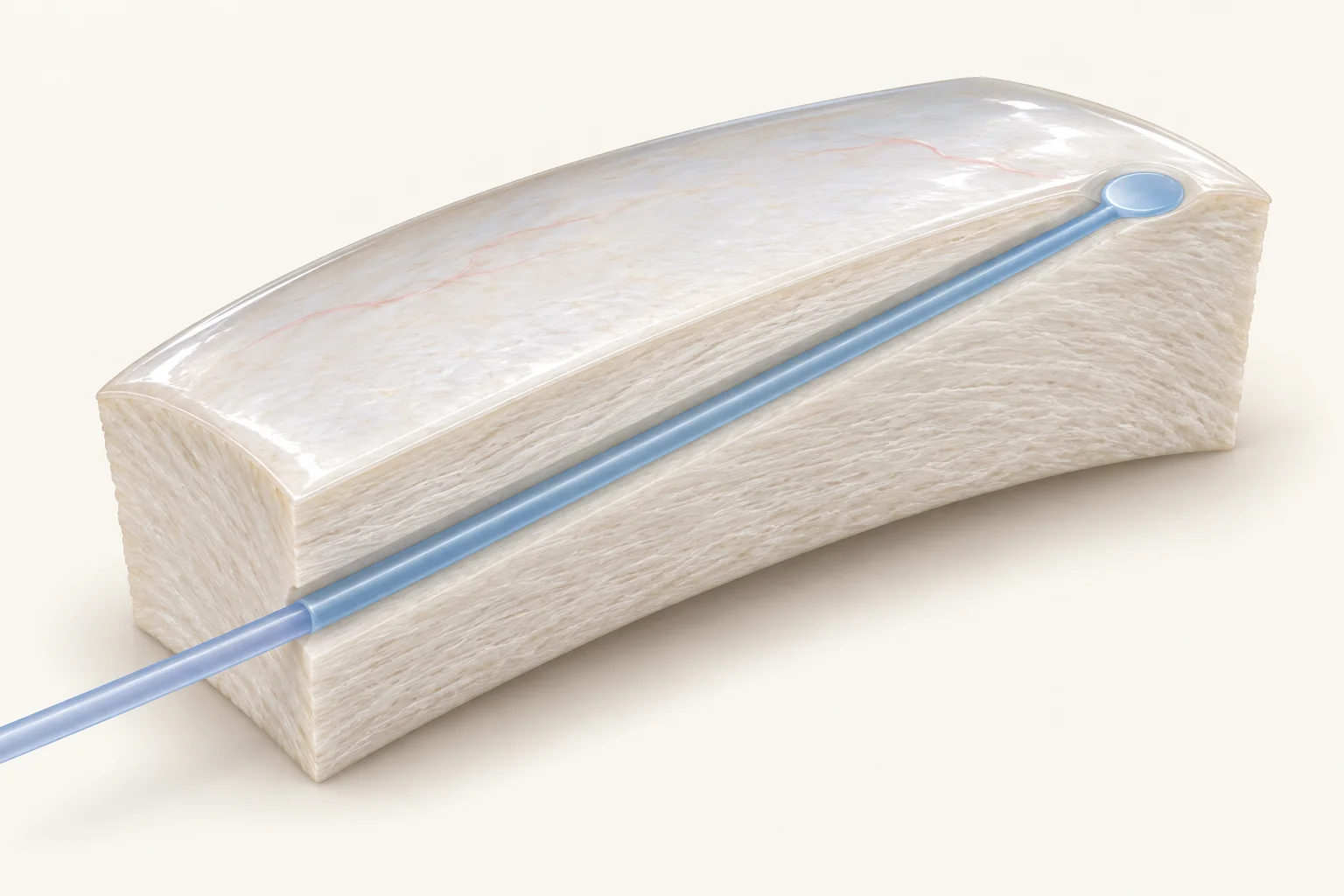
- Conjunctiva (a thin membrane covering the wall of the eye)
- Sclera (the white wall of the eye)
- Flange embedded in the sclera, flush with the surface
- Supporting arm of the lens implant
The surgeon positions the lens behind the iris and uses fine needles to guide its arms through small angled tunnels in the sclera, the white wall of the eye. Controlled heat forms a tiny flange on each tip. The flanges are seated in the scleral tunnels beneath the eye’s surface covering, helping prevent the arms slipping back inside. The lens is not attached to the retina.
The technique is named after Japanese ophthalmologist Shin Yamane. His team published the flanged double-needle method in 2017. It is a surgical fixation method, not a lens brand or a laser treatment. “Sutureless” describes how the implant is held; a separate surgical wound may still need a stitch.
Can my existing implant be kept?
Sometimes. A displaced implant does not automatically need replacing. The surgeon assesses its design, optical clarity, power and supporting arms, as well as the support that remains inside the eye.
A suitable intact implant may be repositioned and secured. A damaged or unsuitable implant may need exchanging. If enough capsule remains, a lens may be supported by the bag or in the sulcus, a space just behind the iris, sometimes with the optic held by the remaining capsular opening. These options have their own requirements and cannot be used in every eye.
Observation may be reasonable when a small displacement is stable, vision is acceptable and the implant is not causing harm. Progressive movement, troublesome vision or effects on other eye structures may favour surgery. The decision starts with assessment of your eye, before choosing a fixation technique.
How does Yamane compare with other fixation methods?
The main difference is which part of the eye supports the implant. Each method has advantages and limitations; the table is a guide to the discussion, rather than a ranking of operations.
| Method | How the lens is supported | What matters for a patient |
|---|---|---|
| Yamane fixation | Two supporting arms are anchored in scleral tunnels with small flanges. The optic sits behind the iris. | Avoids permanent holding sutures. Accurate positioning and a suitable lens are essential; tilt, arm damage or exposure of an anchoring tip can require further treatment. |
| Sutured scleral fixation | Stitches secure a suitable lens to the white eye wall, behind the iris. | Offers several lens and fixation arrangements. Sutures can remain stable for years, but may loosen, break or become exposed. Risk depends on the material and technique. |
| Iris-claw fixation | A purpose-designed implant clips onto small folds of iris tissue, either behind or in front of the iris. | Requires suitable iris tissue. Potential problems include inflammation, pupil distortion or loss of attachment. |
| Iris-sutured fixation | Stitches attach a suitable lens to the iris, usually with the optic behind it. | May allow an existing lens to be retained. Requires adequate iris tissue; iris irritation, pupil changes and suture problems are considerations. |
| Anterior chamber lens | A purpose-designed lens sits in front of the iris, supported at the outer angle of the front chamber. | Can be suitable in selected eyes. Corneal health, chamber depth and drainage-angle anatomy are especially important. |
| Other sutureless scleral methods | Arms are tucked into scleral tunnels or pockets, or purpose-designed anchors support the lens. Some methods use tissue glue to close covering flaps. | These are different operations, not all versions of Yamane. Suitability, implant availability and evidence vary; the optic is not glued to the retina. |
| Capsular or sulcus support | Remaining natural support holds the lens, sometimes with additional support devices or optic capture. | Worth considering when enough suitable capsule remains. It is not possible when that support is inadequate. |
What does the surgeon assess before recommending surgery?
Assessment considers the remaining capsule, the condition of the iris and white eye wall, the cornea, eye pressure and the retina. Previous surgery and the exact implant design also matter. Measurements help plan lens power when a new implant is needed; retinal imaging may help explain the likely visual benefit.
An eye with damaged iris tissue may need a different solution from one with a healthy iris but a vulnerable cornea. Similarly, a technically well-positioned implant cannot remove the effects of macular disease or optic-nerve damage. This is why the recommendation and expected vision must be individualised.
What happens during surgery and recovery?
Fixation may be combined with removal of a displaced implant or remaining natural lens material. A vitrectomy may also be needed to remove vitreous gel from around the implant and surgical openings, or to manage associated problems at the back of the eye. Read more about vitrectomy surgery.
The anaesthetic and length of the operation depend on the work required and your circumstances. Afterwards, follow the surgeon’s instructions about drops, eye protection, activity and review appointments. Vision can fluctuate while the eye settles, and further treatment may be needed if inflammation, pressure changes or retinal swelling occur.
If a gas bubble is used during associated retinal surgery, specific positioning and travel restrictions may apply. Do not fly while gas remains in the eye. Gas is not an automatic part of Yamane fixation; your team should explain whether it has been used.
What are the risks?
Yamane fixation shares risks with other complex lens operations and has some specific to its anchoring method. These include:
- Lens positioning problems: the implant can tilt, move off centre or become caught against the iris, affecting vision.
- Problems with an arm or flange: damage, loosening or exposure through the surface tissue may need repair or another operation.
- Inflammation, bleeding or pressure changes: these may need additional treatment and review.
- Swelling of the central retina: cystoid macular oedema can blur vision and delay recovery.
- Corneal damage: injury to the cells that keep the cornea clear can affect sight.
- Serious complications: retinal detachment or infection inside the eye can threaten vision.
Your personal risk depends on the eye’s condition and the whole operation being performed. A complication percentage from one published series may not describe your situation accurately.
When should I seek urgent help after surgery?
Contact your treating team urgently for increasing pain, worsening redness, a sudden drop in vision, new flashes or a sudden increase in floaters, or a curtain or shadow across your sight. If the team is unavailable, seek urgent assessment at an emergency department. Do not wait for a routine appointment or a reply to an online booking request.
What vision can I expect afterwards?
The aim is a stable lens in a useful optical position. How much clearer you see also depends on the retina, optic nerve and cornea, and whether the implant was the main cause of the visual problem.
Glasses may still be needed. Planning the lens power and final focus can be more complex when a lens sits outside the normal capsular bag, and a good result does not necessarily mean reading or driving without glasses. The intended focus and likely need for spectacles should be discussed before surgery.
For the difference between how a lens focuses and how it is supported, see intraocular lens options.
What else do patients ask?
Will the implant last for life?
It is intended to remain in the eye long term, but no fixation method can be promised to last for life. Yamane studies include follow-up over several years; long-term comparative evidence remains limited. Attend the recommended reviews, even after vision has improved.
Is Yamane the same as laser treatment after cataract surgery?
No. Yamane surgically secures the implant. YAG laser capsulotomy treats clouding of the capsule behind an implant; it does not reattach a loose lens. If an implant may be unstable, its support should be assessed before treatment is planned.
What should I bring to the consultation?
Bring your referral, medication list and any previous operation reports or lens implant details you have. Useful questions include whether the current lens can be retained, why a particular method is recommended, whether vitrectomy is needed, and what is likely to limit the final vision. See planning your visit.
For referring optometrists and ophthalmologists
What should referring clinicians know?
The original flanged double-needle series was prospective but non-comparative. The 2024 Yamane-versus-sutured meta-analysis included 737 eyes in 13 studies; 10 studies were retrospective and only one was randomised. Final BCVA was assessed at 3–12 months. Its findings support Yamane as an option, rather than establishing equivalence or superiority across devices and patient groups.
The 2025 multicentre CME signal should be considered alongside baseline macular status, case selection and postoperative treatment differences. The study’s reported percentage and event denominator are internally inconsistent, so its exact rate should not be presented as a general counselling figure.
Refraction, centration and corrected acuity are distinct outcomes. In the retrospective iris-claw comparison, corrected vision improved with both methods, while refractive predictability differed. Lens design, haptic behaviour, fixation geometry and the surgeon’s own outcomes deserve attention; a formula, lens constant or device preference should not be transferred uncritically between techniques.
Simon Chen’s 2020 technical article on Yamane fixation provides additional surgical context. It is a dated expert discussion, rather than a comparative trial. Current referral information is available separately.
References and further reading
- Yamane S et al. Flanged intrascleral intraocular lens fixation with double-needle technique . Ophthalmology. 2017.
- Shen JF et al. IOL implantation in the absence of zonular support: AAO outcomes and safety update . Ophthalmology. 2020.
- Yamane versus sutured scleral fixation: systematic review and meta-analysis . J Clin Med. 2024.
- Ishikawa H et al. Cystoid macular oedema after Yamane fixation: multicentre cohort study . Sci Rep. 2025.
- Retropupillary iris-claw versus flanged intrascleral fixation . J Cataract Refract Surg. 2024.
- Long-term visual outcome and clinical predictors following Yamane fixation . 2026.
- Chen S. Pearls for the Yamane technique . CRST Europe. February 2020.
- American Academy of Ophthalmology EyeWiki. Scleral-fixated intraocular lenses .
- American Academy of Ophthalmology EyeWiki. Anterior chamber intraocular lenses .
- Guy’s and St Thomas’ NHS Foundation Trust. Vitrectomy: patient information .
- American Society of Retina Specialists. Intraocular lens dislocation . Patient information; 2016.
These are the published sources behind this article. They are written for clinicians and do not replace advice about your own eye.
How do I make an appointment with Dr Chen?
Appointments and enquiries through Vision Eye Institute, Chatswood.
Clinic appointments: (02) 9424 9999