
Flashes, floaters or a curtain in your vision: when is it urgent?Flashes, floaters or a shadow: what should I do?
A new curtain, shadow or missing area of vision needs immediate advice from an optometrist, ophthalmologist or emergency eye service. For new flashes or floaters, contact an eye-care professional promptly to arrange assessment, even if your sight otherwise seems clear and your eye does not hurt.
These symptoms often come from a change in the gel inside the eye. Sometimes, however, that change tears the retina. You cannot reliably tell the difference from the symptoms alone.

What do flashes, floaters and a curtain look like?
Does a vitreous detachment mean my retina has detached?
No. The vitreous is the clear gel that fills much of the eye. The retina is the thin, light-sensitive lining at the back, rather like the sensor in a camera.
With age, the gel changes and can separate from the retina. This is called a posterior vitreous detachment, or PVD. An uncomplicated PVD leaves the retina attached and usually does not need treatment.
Sometimes the separating gel pulls hard enough to create a retinal tear. Fluid can then pass through the tear and lift the retina away from the eye wall, causing a retinal detachment. This is why an apparently simple new floater needs checking.
Partial vitreous detachment
Retinal tear and detachment
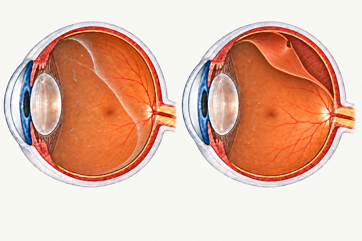
Short-sightedness, an eye injury, previous cataract surgery and a previous retinal detachment can increase concern. Tell the examiner about these—but remember that new symptoms need assessment even without a known risk factor.
What happens during the eye examination?
The examiner checks your sight and looks for a tear, bleeding or detachment. Drops usually widen the pupil so that the retina can be examined, including its outer edges.
A careful peripheral retinal examination may involve a special lens and gentle pressure around the eye. This can feel uncomfortable. Tell the examiner if you are struggling so they can help you through it.
If the facility has ultra-widefield retinal imaging, using a camera such as an Optos device, this can photograph a large area of the peripheral retina and can clearly show retinal tears or a retinal detachment. It is a useful addition to the examination, but some tears can be missed, so a normal image does not rule one out. Read more about retinal imaging technology.
Scans can also add information. Ultrasound may be useful when bleeding or another obstruction makes the retina difficult to see. An OCT scan gives detailed images of retinal layers, but a normal central OCT image alone does not rule out a tear at the edge of the retina.
If there is a tear, laser or freezing treatment may be recommended to reduce the chance of detachment. A detached retina often needs surgery. The examination determines which advice applies; flashes and floaters alone do not establish a diagnosis or a need for treatment.
Can a tear develop after a reassuring first examination?
Yes. When a PVD starts, the vitreous does not necessarily separate from the whole retina in one go. Separation is a process that progresses over a variable period, sometimes taking months. As it continues, the gel can pull on areas where it is still attached and cause a new tear.
The risk of a retinal tear or detachment is generally highest when symptoms begin and during the first six weeks. After that, the risk diminishes but does not disappear. A reassuring examination describes what was seen at that visit; new symptoms still need attention later.
The period of greatest risk as the vitreous separates.
Risk diminishes, but new symptoms still need assessment.
Follow the review plan your examiner gives you. Timing depends on the findings and your risk factors, so there is no single follow-up interval suitable for everyone.
Seek urgent reassessment for more floaters, new or increasing flashes, reduced vision, or any new shadow or curtain. Do not wait for your booked follow-up. This also applies if you have already had treatment for a tear: a separate new tear can occur.
If you cannot remember the return advice, contact the service that examined you. Keep their instructions somewhere easy to find.
Could the lights be migraine?
The flashing lights of migraine usually have a different pattern from those of a PVD.
| Feature | PVD flashes | Migraine visual aura |
|---|---|---|
| Which eye? | Usually one eye at a time | Generally both eyes at the same time |
| How long? | A split-second flash, which may recur | An evolving disturbance lasting 5–30 minutes, sometimes longer; typically up to 60 minutes |
| Appearance | A brief white arc, lightning-like flicker or camera flash at the outer edge of vision | White or multicoloured shimmering lights, often zigzag-shaped; sometimes around a blurred or missing spot |
| Lighting | Most noticeable in low light or darkness | Often apparent in normal lighting as well |
| Other features | May follow rapid head or eye movement and accompany new floaters | May be followed by a headache, but can occur without one |
These patterns help the clinician, but are not a reliable home test. Do not assume a first, different or unexplained episode is migraine, particularly if vision is suddenly lost or part of your view is missing. Seek prompt assessment even if that loss of vision clears.
What if my floaters are longstanding and troublesome?
Once urgent retinal problems have been excluded, persistent floaters can be discussed at a routine appointment. Their effect on reading, driving or daily activities matters. That is a separate decision from checking a new symptom.
For floaters that remain troublesome after an eye examination, read about YAG laser for selected floaters, vitrectomy surgery, and recovery after vitrectomy for floaters. These treatment decisions are separate from the prompt assessment of new flashes, floaters or a shadow.
What information helps an urgent referral?
For GPs and optometrists: document onset, laterality, change in floaters or flashes, visual loss or field symptoms, measured acuity in each eye, relevant trauma or surgery and previous retinal disease. Make prompt clinical contact for acute symptoms, with a view to assessment today or within the timeframe advised by the eye-care professional. Suspected detachment or sudden visual loss needs immediate consultation.
Direct ophthalmoscopy alone cannot exclude a peripheral tear. Arrange a direct clinical handover where possible, and confirm where the patient should attend. Routine electronic referrals or booking forms should not be relied upon for emergency triage.
What should I do now?
- Seek advice immediately: for a new curtain, dark shadow, missing area of vision or sudden sight loss, consult an optometrist, ophthalmologist or emergency eye service immediately. Explain that you have new visual loss so they can arrange urgent assessment. If you cannot reach an eye-care professional promptly, attend an emergency eye service.
- Make contact promptly: for new flashes, new floaters or a sudden increase in floaters, contact an eye-care professional with a view to being seen today, or within a short timeframe based on that professional's recommendation. Do not decide to wait several days without discussing the symptoms.
- Arrange routine review: for longstanding floaters that have not changed. If they have never been examined, arrange an eye examination. Any new change needs prompt advice.
A retinal tear can occur without a curtain, and a detachment may begin at the edge of your vision while reading vision remains clear. Do not wait for pain or worse sight before making contact. A routine booking form or online message is not a reliable way to obtain urgent advice.
Sources and further reading
- National Eye Institute: retinal detachment.
- American Society of Retina Specialists: posterior vitreous detachment and retinal tears.
- healthdirect: eye floaters.
- RACGP: flashes and floaters—assessment and management.
- Nixon, Davie and Snead: prospective study of PVD and retinal tears. Eye, 2024.
- RNIB: understanding posterior vitreous detachment.
- International Headache Society: migraine with aura.
- Chou and colleagues: ultra-widefield imaging for retinal breaks. Eye, 2026.