
Can a lens implant be replaced after cataract surgery?
IOL exchange explained
Yes. A lens implant can often be removed and replaced after cataract surgery, even many years later. This operation is called an intraocular lens exchange, or IOL exchange. Whether it is worth doing depends on what is causing the problem, how much it affects your vision, the condition of the eye and the risks of another operation.

Most lens implants are intended to remain in the eye for life. They do not need routine replacement. If a problem develops, the existing lens may sometimes be repositioned, or the symptoms treated without exchanging it.
Why might a lens implant need to be replaced?
There are several reasons. Some arise soon after cataract surgery; others develop years later. Establishing the cause matters because similar symptoms can need very different treatments.
Is the lens power or glasses prescription different from expected?
A refractive surprise means the eye is more short-sighted, long-sighted or astigmatic after surgery than intended. The two eyes may also have an uncomfortable difference in focus.
This can result from limitations in eye measurements and lens-power predictions, previous corneal surgery, the implant's final position, or occasionally implantation of an unintended lens power. An unexpected result does not necessarily mean that the wrong lens was inserted.
An exchange can change the implant's power, but it is only one option. Glasses, contact lenses, selected corneal laser treatment or a purpose-designed supplementary lens may be suitable. The prescription and eye surface need assessment before deciding which approach makes sense.
Has a toric lens rotated or another implant moved?
A toric implant corrects astigmatism and needs to sit at the planned angle. If it rotates, its effect changes. When the lens power is appropriate and the support is sound, rotating the existing implant may be preferable to replacing it.
Other implants can tilt, move away from the centre or slip from their normal position. Partial displacement is called subluxation; more complete displacement is called dislocation. Possible symptoms include blurred or fluctuating vision, glare or seeing the edge of the lens.
The implant normally sits inside the thin capsule left behind when the cataract is removed. Fine fibres called zonules suspend this capsular bag. Injury, previous surgery or conditions that weaken these fibres, such as pseudoexfoliation, can allow the lens and sometimes the whole bag to move. An intact implant may be secured again; a damaged or unsuitable one may need exchange.
The capsular bag and its supporting fibres help hold the implant in place. This simplified illustration omits the iris and cornea so the support is visible.
Can the artificial lens itself become cloudy?
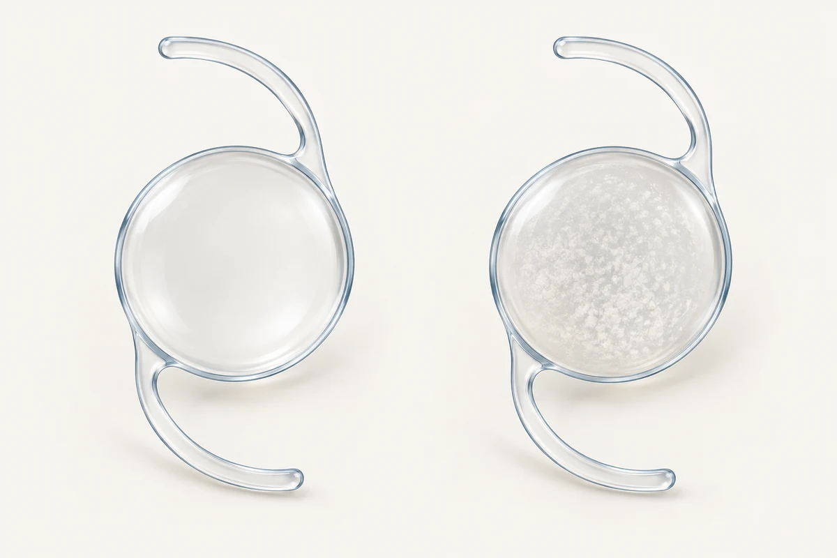
Yes. Some implants develop deposits or changes in their material that reduce transparency. This is called IOL opacification. It is different from a cataract returning: the original natural lens has already been removed.
Certain older Oculentis LENTIS lenses have been affected by calcification. An Australian recall notice in 2014 concerned specified lenses supplied in glass vials. This does not mean that every Oculentis lens will become cloudy or should be removed. The exact model, its appearance and its effect on vision need checking.
Clouding can also develop in susceptible hydrophilic acrylic implants after some operations involving air or gas inside the eye. These include certain corneal endothelial transplants and retinal operations using vitrectomy and gas. Calcium deposits have been identified in affected lenses. The material and circumstances matter; this is not an inevitable consequence of having gas in the eye.
Silicone oil is a different issue. Oil used in some retinal operations can adhere to an implant's surface, particularly a silicone implant, and interfere with its optical clarity. This is distinct from the calcium deposits associated with hydrophilic lenses. Assessment determines whether cleaning, exchange or another approach is appropriate.
A clear implant and an illustration of deposits within a cloudy implant. These are generic examples, not photographs or depictions of a particular commercial model.
Could the implant be rubbing on the iris?
An implant in an unsuitable position, or a design unsuitable for that position, can rub against the iris. This may cause recurrent iritis or uveitis (inflammation), pigment release, bleeding or raised eye pressure. The combination of inflammation, pressure problems and bleeding is sometimes called uveitis–glaucoma–hyphaema syndrome, or UGH syndrome.
For example, a single-piece acrylic implant designed for the capsular bag can cause problems if placed in the sulcus, the space just behind the iris. However, sulcus placement itself is not necessarily incorrect: an appropriately designed lens can be placed there deliberately when sufficient support remains. Repositioning or exchange may be considered if the implant is causing ongoing irritation.
Can YAG laser damage a lens implant?
A YAG laser capsulotomy can occasionally leave small pits in the implant. Their effect depends on their number, size and position. A small pit does not automatically require treatment; central or more extensive damage may contribute to glare or poorer visual quality. Laboratory studies demonstrate that pits can scatter light, but they cannot tell an individual patient whether exchange will help.
The surgeon needs to establish whether the marks explain the symptoms and weigh the potential benefit of replacement against its risks.
What if vision is sharp on the chart but the quality is poor?
Some people remain troubled by haloes, glare, starbursts, reduced contrast or a waxy quality of vision after a multifocal or extended depth-of-focus (EDOF) implant. The symptoms and trade-offs differ between lens designs.
Before blaming the implant, assessment should look for a residual prescription, dry eye, a cloudy capsule, lens decentration and corneal or retinal disease. Treating another cause may improve vision without exchange.
If troublesome symptoms persist and are attributed to the lens optics, exchange to a different design may be considered. Moving from a multifocal or EDOF implant to a monofocal lens usually means accepting more dependence on glasses, particularly for reading. It does not guarantee that every visual symptom will disappear. Evidence from older multifocal exchange studies should not be treated as a success rate for every modern EDOF lens.
Negative dysphotopsia is another possible reason for intervention. It describes a dark crescent or shadow towards the outer side of the vision. If persistent and troublesome, it may respond to changing how the implant sits relative to the front of the capsule. Options can include repositioning, changing the relationship between the optic and capsule, a supplementary lens or exchange. Simply swapping one lens for another in the same position may not resolve it.
Other less common reasons for exchange include a damaged implant or broken supporting arm. The need for surgery depends on its effect on the eye, not just the presence of an abnormal finding.
Could the cloudy area be behind the implant instead?
Yes. Posterior capsule opacification (PCO) is clouding of the thin membrane behind the implant. It is different from clouding within the artificial lens itself.
When PCO is responsible for reduced vision, a YAG laser capsulotomy can make an opening in that membrane. It does not remove calcium deposits from a cloudy implant or correct an unsuitable lens power.
If exchange is being considered, discuss that possibility before proceeding with YAG treatment. Opening the posterior capsule changes the support available and can make a later exchange more complex. A previous capsulotomy does not make exchange impossible, but it may change the surgical plan or the need to remove vitreous gel that comes forward. Studies report differing complication rates in different patient groups, so there is no single risk figure that applies to everyone.
How is a replacement lens held in place?
The replacement does not always have to sit in exactly the same place as the original. The surgeon assesses the remaining capsule, zonules, iris, cornea and white wall of the eye, called the sclera. Several approaches are possible.
- Inside the capsular bag: if the bag and its supporting fibres remain usable, a replacement can sometimes be placed in the original position.
- In the sulcus: a suitable lens may sit just behind the iris when enough capsular support remains. In selected eyes, the optic can also be held by the capsule opening, a method called optic capture.
- Attached to the iris: selected implants can be secured with sutures or an iris-claw design. This requires suitable iris tissue.
- In the anterior chamber: a specially designed lens can sit in front of the iris. The health of the cornea, drainage angle and other structures influences suitability.
- Fixed to the sclera: the lens is supported by the eye wall using sutures or a sutureless technique, rather than relying on the original capsular bag.
The Yamane technique is one form of sutureless scleral fixation. The supporting arms of a suitable lens, called haptics, are secured in scleral tunnels with small flanged ends. It is an option for selected eyes without adequate capsular support.
Hoffman pockets are different: small pockets in the sclera cover and protect fixation sutures and their knots. This is a way of performing sutured fixation, rather than another name for the Yamane technique. Other sutured and sutureless methods are also available.
No single method is best for every eye. Each has its own requirements and possible complications. A previously used multifocal or toric design may not be suitable for the new position, and a different lens type or focusing target may be needed.
What happens during lens exchange surgery?
The surgeon frees the original implant from surrounding tissue and removes it through an incision. Depending on its material and design, it may be folded, divided or removed through a larger opening. The replacement is then positioned using the most suitable support.
A vitrectomy, which removes some of the eye's vitreous gel, may be needed if the gel has come forward, the lens has fallen backwards or a retinal procedure is required. Occasionally the safest plan involves removing the implant first and placing a replacement in a separate operation. These possibilities should form part of the discussion beforehand.
The anaesthetic, day-surgery arrangements and recovery plan depend on the operation required. Recovery after a straightforward exchange can differ substantially from recovery after an exchange combined with retinal or corneal surgery.
Is it too late to change a lens years after cataract surgery?
Not necessarily. Exchange can be performed years after implantation. However, the capsule may have contracted or become firmly attached to the implant, and the supporting fibres or other eye structures may have changed. These factors can make removal more involved.
There is no universal time limit. It is sensible to seek assessment when a problem is affecting daily life, rather than assuming it is either too early or too late. Some early symptoms need time or treatment of another cause; a displaced lens or ongoing damage to eye structures may require a more prompt decision.
What are the risks and likely benefits?
The aim is to address a defined problem: for example, improve focus, remove a cloudy optic, stabilise a displaced lens or stop irritation. The amount of improvement also depends on the cornea, retina and optic nerve. Replacing the implant cannot reverse unrelated damage to those structures.
Possible complications include damage to the capsule or its supports, bleeding, infection, persistent inflammation, swelling at the macula, raised eye pressure, corneal damage, retinal tears or detachment, and movement or tilt of the replacement lens. A further operation may be needed. Serious complications can cause permanent loss of vision.
The new prescription can still differ from the intended result. Glasses may remain necessary, and glare or other symptoms may persist. Published outcomes come from very different groups of patients and operations, so an overall success percentage is less useful than an assessment of your particular eye.
If symptoms are tolerable, vision is adequately corrected and the implant is not harming the eye, observation may be the most appropriate choice.
What should I bring to an assessment?
Bring your implant card or lens details if available, the date and records of your cataract surgery, your current glasses and details of any later YAG laser, retinal or corneal treatment. Explain what is difficult in daily life: reading, driving at night, fluctuating focus, glare or a shadow can point to different problems.
Useful questions include:
- Is the implant itself the main cause of my symptoms?
- Could treatment, glasses or repositioning help without an exchange?
- What support remains, and where would a replacement sit?
- What improvement is realistic, and which symptoms may remain?
- What are the particular risks in my eye, and what is the alternative if I wait?
Seek urgent eye assessment for sudden loss of vision, new flashes, a sudden increase in floaters or a curtain-like shadow. A painful red eye also needs prompt assessment. Do not wait for a routine lens-exchange consultation if these symptoms occur.
For a planned assessment with Dr Chen, contact Vision Eye Institute Chatswood through the appointments page. You can also read about IOL exchange and complex cataract surgery and lens implant options.
References and further reading
- Patel and colleagues. IOL exchange: indications, outcomes and complications . 2023.
- Son and colleagues. Visual outcomes and complications after IOL exchange: IRIS Registry analysis . 2024.
- Kaiser and colleagues. IOL exchange with an open posterior capsule . Published online 2024; journal issue 2025.
- Alsetri and colleagues. Exchange with an open or intact posterior capsule . 2023.
- Kristianslund and colleagues. Randomised comparison of repositioning and exchange for late lens dislocation . 2017.
- American Academy of Ophthalmology. Lens implantation without zonular support: evidence assessment . 2020.
- Yamane and colleagues. Flanged intrascleral lens fixation . 2017.
- Hoffman, Fine and Packer. Scleral fixation without conjunctival dissection . 2006.
- Masket and colleagues. Surgical management of negative dysphotopsia . 2018.
- Kamiya and colleagues. Multifocal lens explantation: a series of 50 eyes . 2014.
- Woodward and colleagues. Dissatisfaction after multifocal lens implantation . 2009.
- Hu. Repositioning of toric implants after rotation . 2023.
- Marcovich and colleagues. Hydrophilic lens opacification after vitrectomy and gas . 2018.
- Therapeutic Goods Administration. Historical Oculentis LENTIS recall notice, 17 December 2014 . Notice reproduced in a 2026 FOI release.
- Kirk and colleagues. Pathological findings with asymmetric or sulcus lens fixation . 2012.
- Dick and colleagues. Silicone oil interactions with implant materials . 1997; laboratory study.
- Borkenstein and colleagues. Effect of YAG laser pits on lens optical quality . 2022; laboratory study.
- Fernández-Buenaga and colleagues. Exchange, supplementary lens or LASIK for residual refractive error . 2013.
- National Eye Institute. Retinal detachment: symptoms and urgent action . Updated 2025.
These are the published sources behind this article. They are written for clinicians and do not replace advice about your own eye.
How do I make an appointment with Dr Chen?
Appointments and enquiries through Vision Eye Institute, Chatswood.
Clinic appointments: (02) 9424 9999