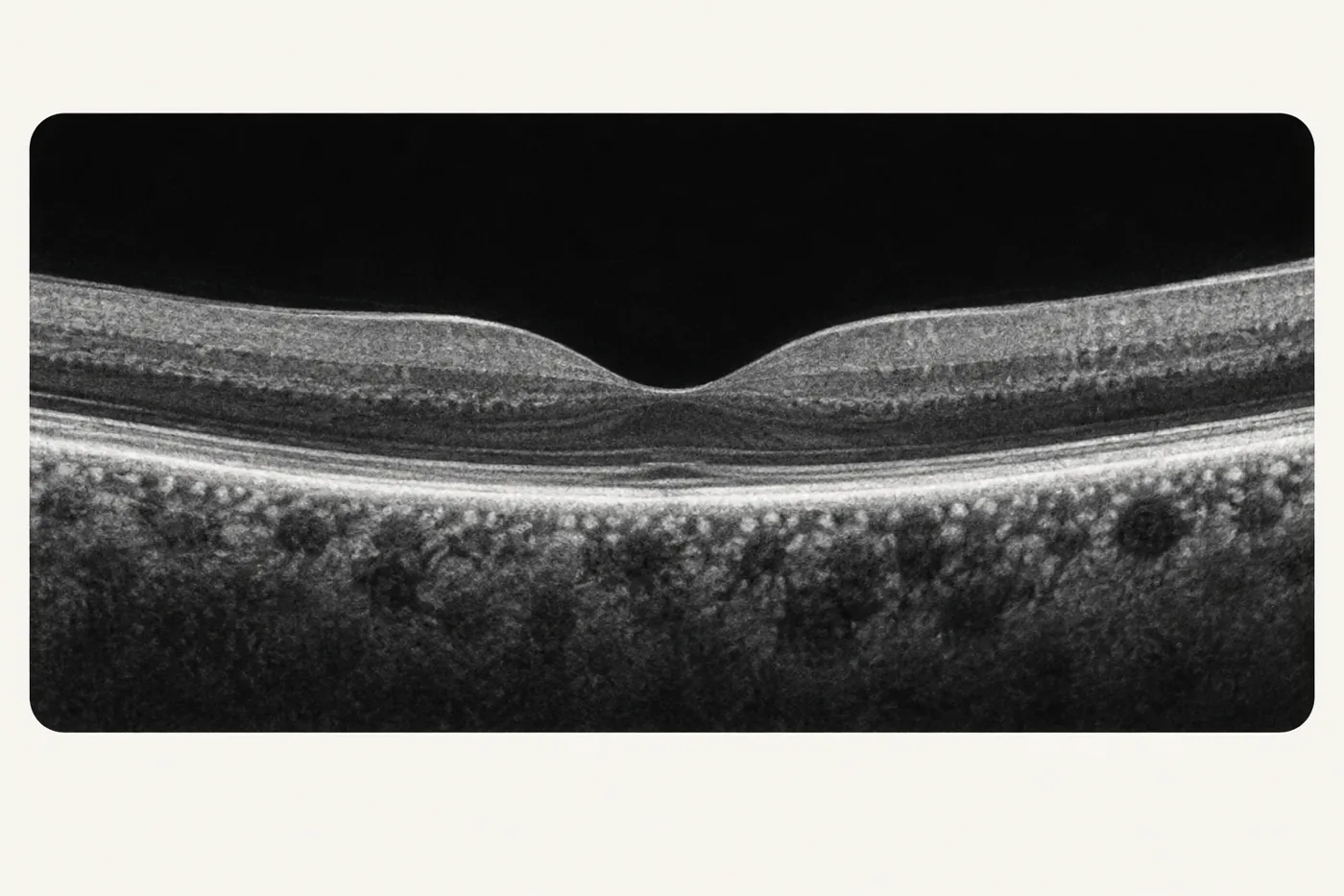
Optical coherence tomography (OCT)
Harmless light waves take cross-section pictures of the retina, a little like an ultrasound scan but with light instead of sound.
 Dr Simon ChenCataract & Retina Surgeon
All of the technology on this page is used by and available to Dr Chen at Vision Eye Institute Chatswood. These advanced, state-of-the-art imaging, laser and surgical systems support his cataract and retinal care. Each has a specific job: to measure the eye accurately, to see the retina in detail, or to make surgery more controlled.
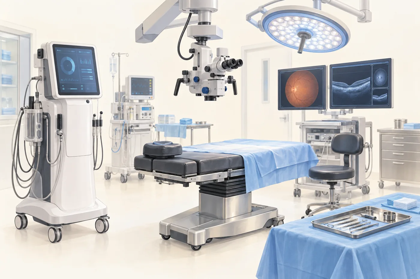
Dr Chen’s clinic uses ultra-widefield retinal imaging, optical coherence tomography (OCT), OCT angiography (OCTA), ultrasound and visual field testing. Together they show the whole retina, its layers and its blood vessels.
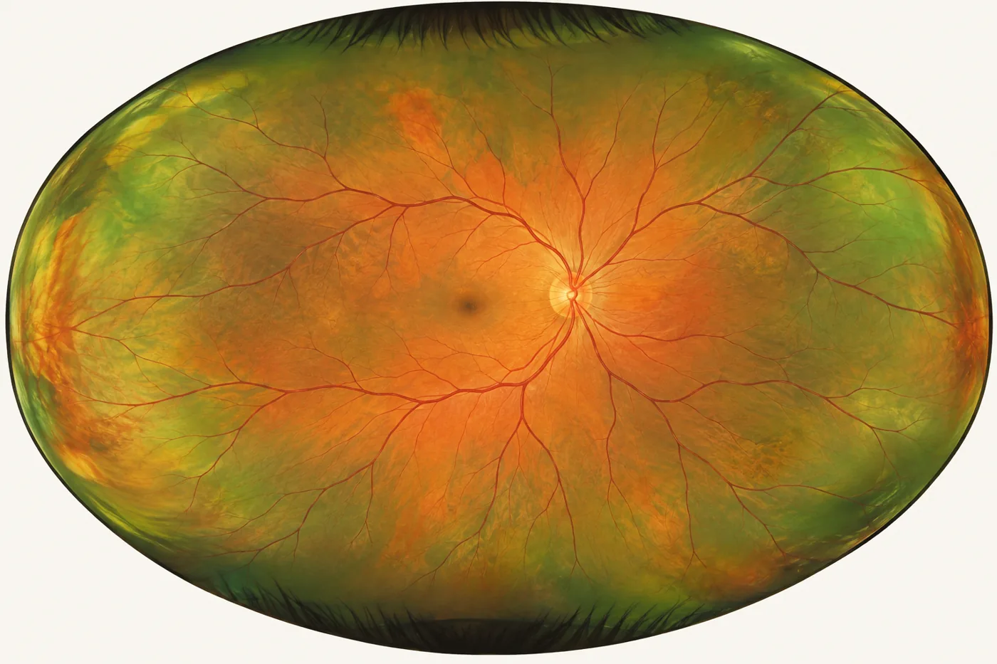
Photographs most of the retina in one image, including the far edge where tears and detachments often begin.
Harmless light waves take cross-section pictures of the retina, a little like an ultrasound scan but with light instead of sound.
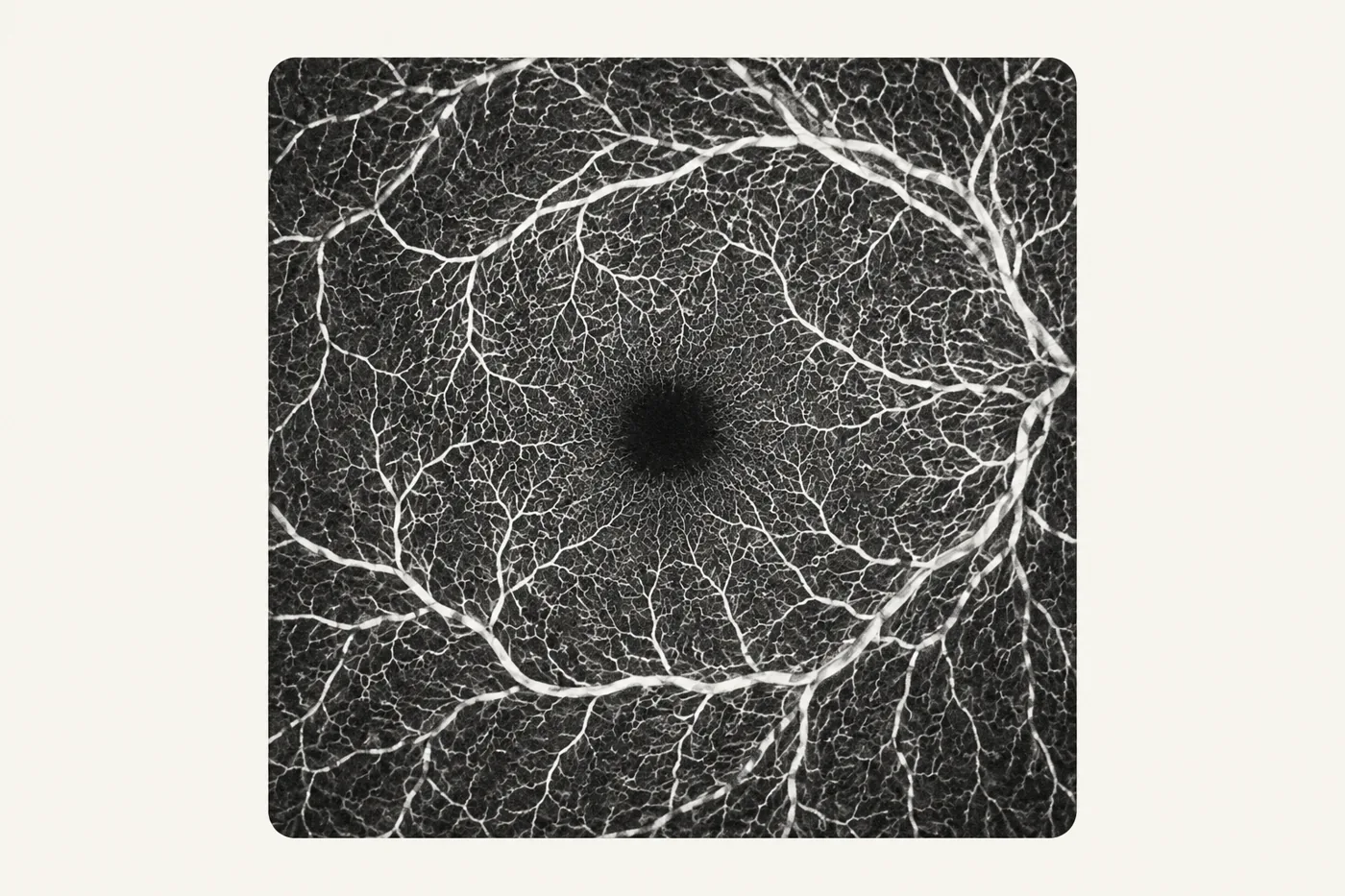
The same scanner maps the retina’s small blood vessels without an injection of dye, which helps with macular degeneration and vein occlusions.
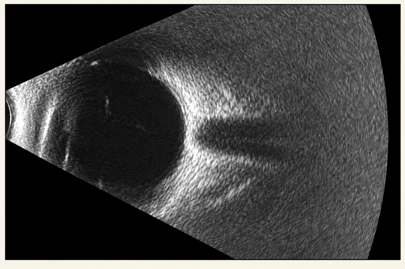
Looks inside the eye when bleeding or a dense cataract blocks the view, to check whether the retina is attached.
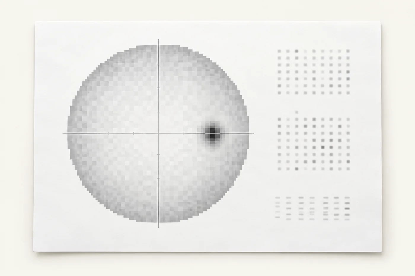
Measures side vision when glaucoma or another condition may also be affecting the eye.
Your eye is measured with optical biometry, multiple methods of corneal mapping and specular microscopy, so the lens power and surgical plan rest on cross-checked measurements.
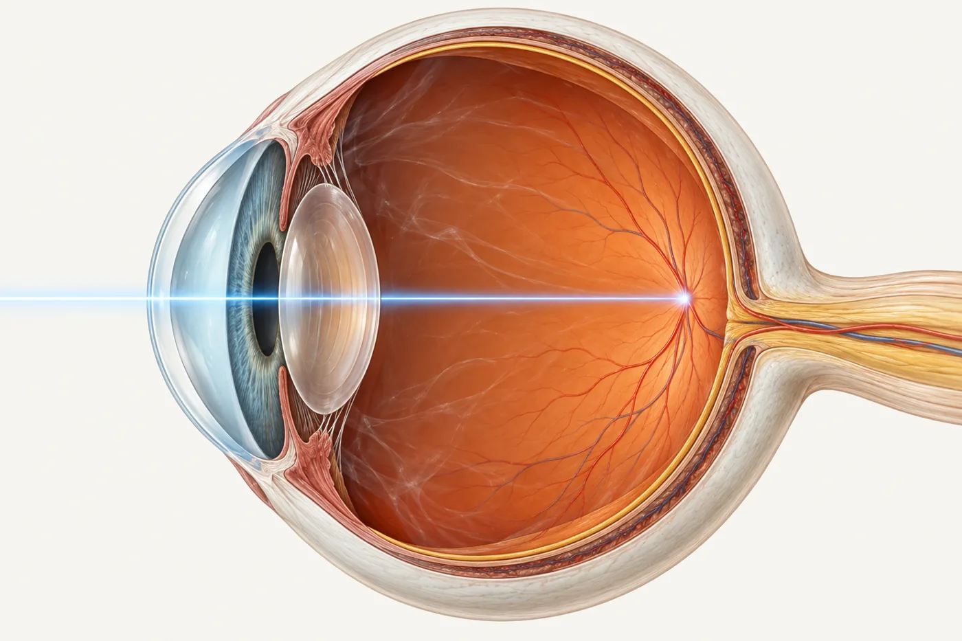
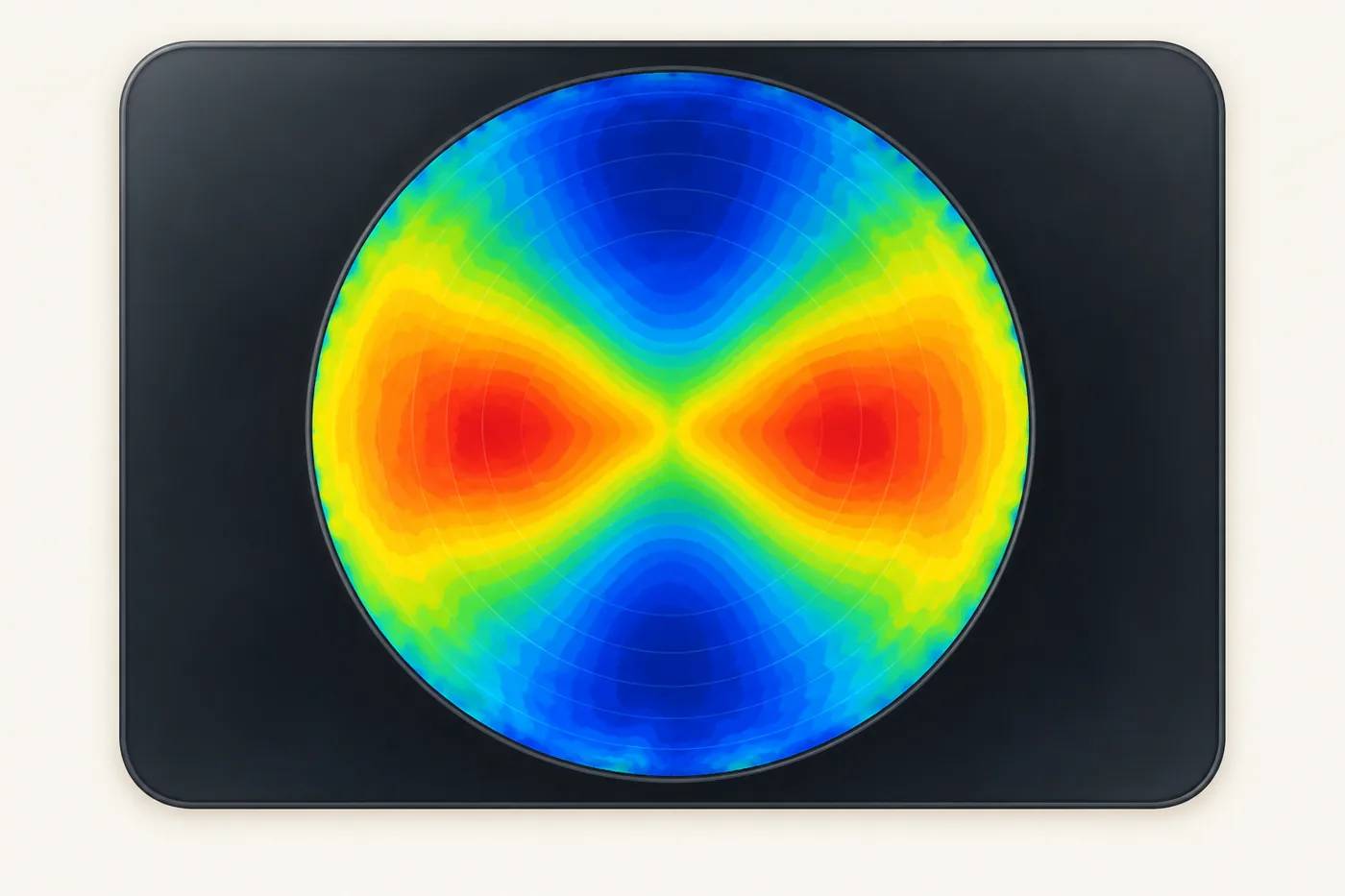
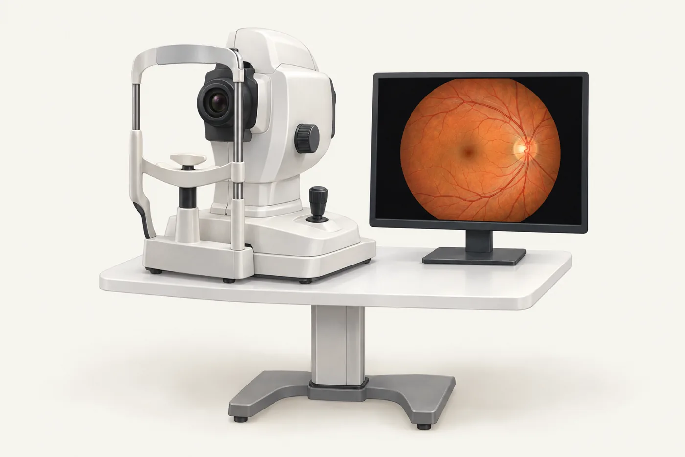
Measures the length and shape of the eye to calculate the power of the lens implant. Its swept-source OCT can measure through dense cataracts, and it shows an image of the centre of the retina to confirm you were looking straight at the target. It also measures the back surface of the cornea, which improves astigmatism planning.
Reads the reflection of illuminated rings from the front of the cornea to map its shape. It also checks the tear film, which must be healthy for accurate measurements.
A rotating camera images the cornea in three dimensions, front and back, and measures its thickness. It helps detect keratoconus and plan surgery after laser eye surgery.
Measures how light is distorted through the whole eye, separating the cornea from the inside of the eye, and measures pupil size in different lighting. This helps decide who is likely to see well with an extended depth of focus or multifocal lens.
Counts the cells lining the back of the cornea. These cells keep the cornea clear, so their health matters before complex or combined surgery.
Optical coherence tomography of the front of the eye, including the drainage angle and the space behind the iris where some lenses sit. It is used for selected eyes, not routinely before cataract surgery.
Estimates how well the retina could see once a cataract is removed, by projecting letters through clearer parts of the lens. It helps set realistic expectations when a cataract and a retinal condition occur together.
Lens power is calculated with latest-generation formulas from several independent measurements, then checked by an experienced, dedicated team in every case, routine or complex.
Several instruments measure the eye independently, so any result that does not fit can be spotted and repeated.
Lens power is calculated with latest-generation formulas, including specialised methods for eyes that have had previous surgery.
Vision Eye Institute is a long-established cataract and refractive surgery practice, and its team has decades of combined experience in lens calculations. Every calculation is checked.
Dr Chen’s clinic has a pattern scanning retinal laser, a YAG laser and a selective laser trabeculoplasty (SLT) laser.
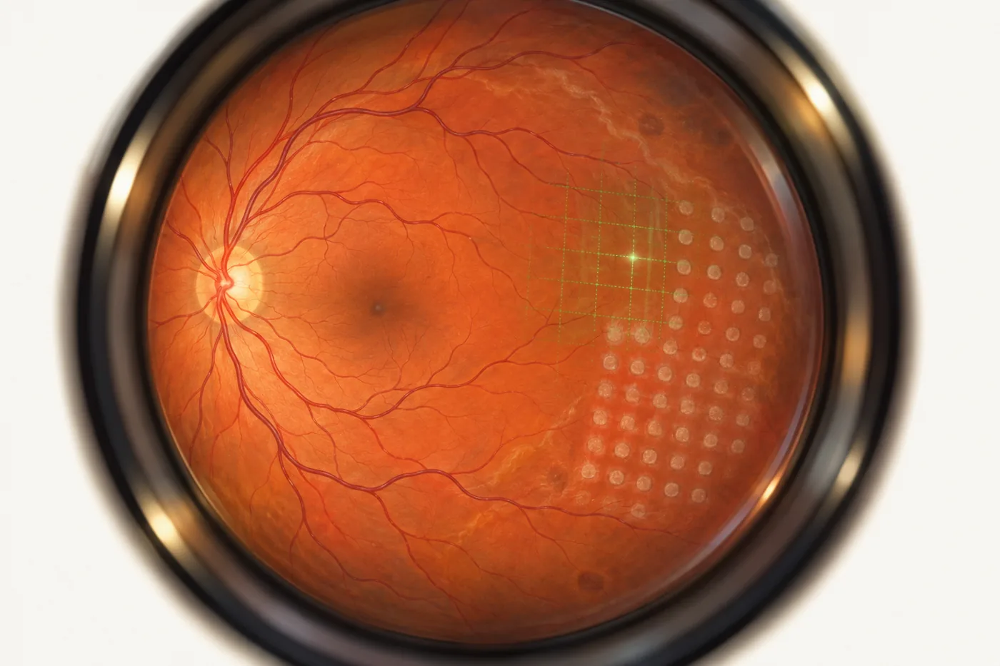
Delivers short pulses in patterns rather than single spots. Treatment is quicker and usually more comfortable than older single-spot laser, with less spread of heat into surrounding tissue.
Clears cloudiness behind a lens implant, makes a small drainage opening in the iris when needed, and is used for YAG laser vitreolysis in selected floaters.
Lowers eye pressure in open-angle glaucoma.
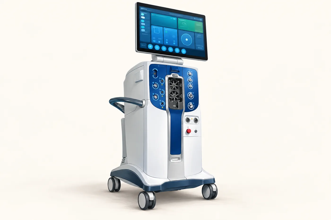
Cataract surgery in Dr Chen’s day surgery uses ZEISS CALLISTO eye digital guidance, the Alcon UNITY Vitreoretinal Cataract System (VCS), a femtosecond laser for selected patients and a high-resolution operating microscope.
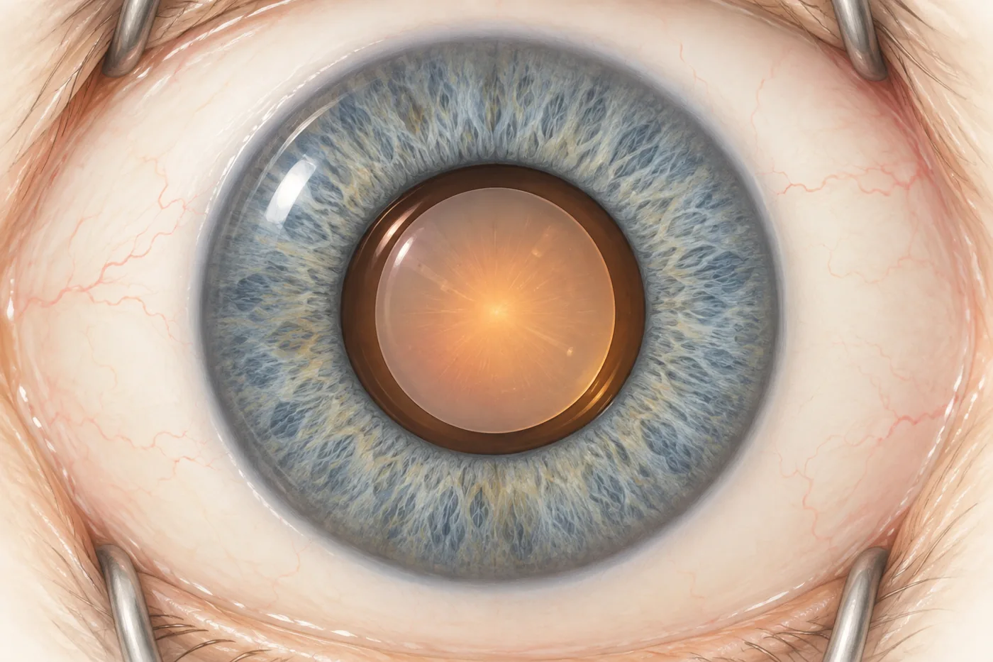
A reference image of your eye is taken when it is measured. In theatre, the system recognises your eye and overlays guides in the microscope: where to place the incisions, the size and position of the capsule opening, and the exact alignment of a toric lens. No ink marks are needed.
The system that removes the cataract.
For selected patients, a laser creates the incisions and a round capsule opening and softens the cataract before it is removed.
Femtosecond laser cataract surgeryA high-resolution surgical microscope with coaxial illumination gives a bright, stable red reflex, the orange glow that shows the lens capsule clearly.
Retinal surgery (vitrectomy) uses the Alcon UNITY Vitreoretinal Cataract System (VCS) with a cutter that works at up to 30,000 cuts per minute, stitchless 23, 25 or 27 gauge instruments, ZEISS RESIGHT wide-angle viewing and laser, freezing, dye and gas or oil treatments inside the eye.
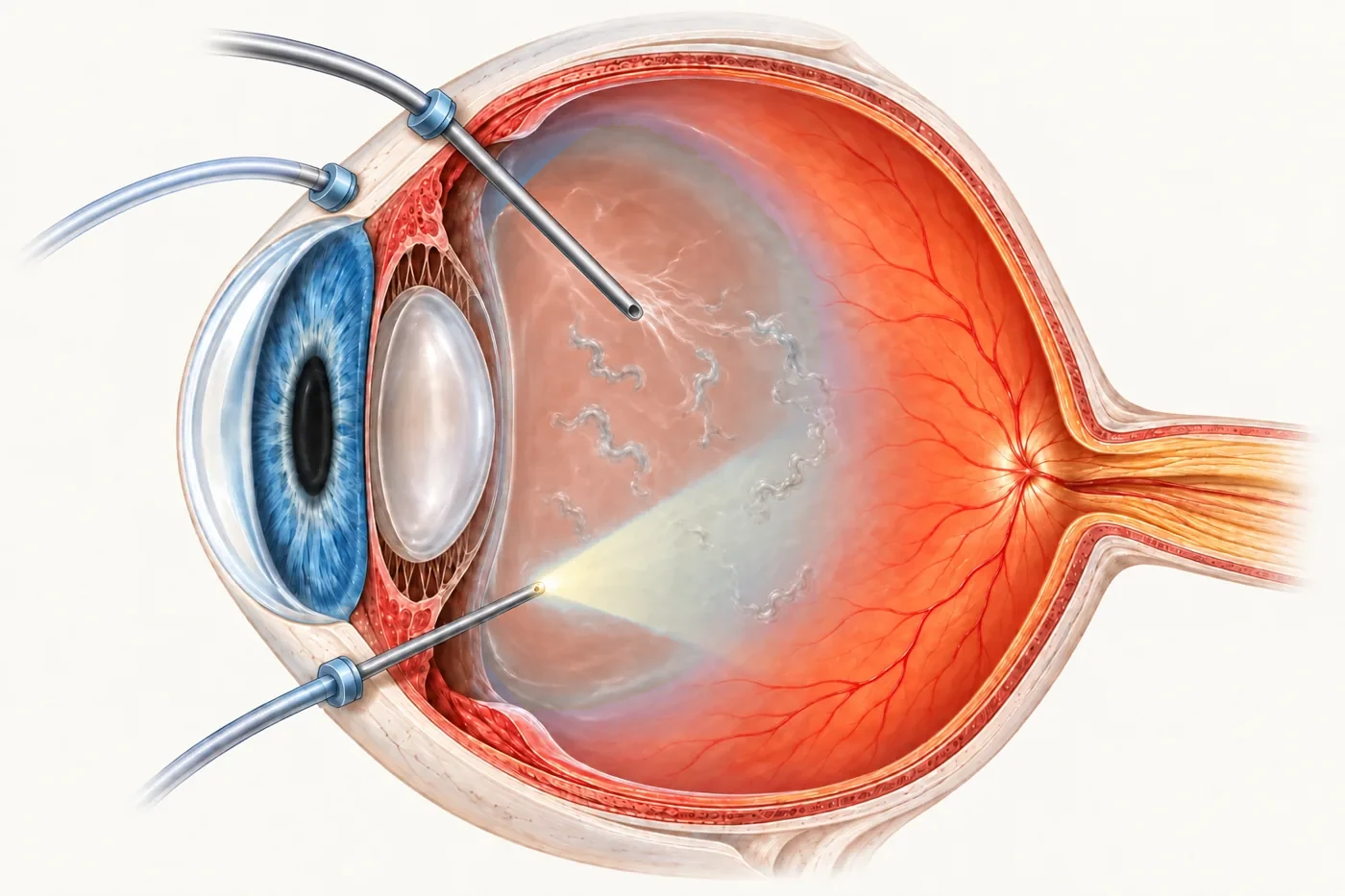
Dr Chen’s vitrectomy platform. Its HyperVit cutter works at up to 30,000 cuts per minute. Faster cutting means each cut takes a smaller bite of the vitreous gel, which is designed to reduce pulling on the retina. The system controls pressure inside the eye throughout, and the same system is used for cataract surgery.
Instruments pass through tiny self-sealing openings of 23, 25 or 27 gauge, about 0.6 to 0.4 mm across. Stitches are rarely needed.
Lets Dr Chen see to the far edge of the retina during surgery, where tears are found and treated.
Curved and illuminated laser probes seal retinal tears. Diathermy controls bleeding. Freezing treatment (cryotherapy) and a head-mounted laser treat tears far forward.
A specialised dual dye makes fine membranes visible for peeling. Heavy liquids, gas and silicone oil flatten and support the retina.
Gas and oil after vitrectomyDr Chen can offer any intraocular lens commercially available in Australia, piggyback lenses, custom-made high-, low- and negative-power lenses, and specialised instruments for lens exchange and scleral fixation.
Dr Chen is not tied to any single manufacturer, so the advice is tailored to your eye.
Lens choicesPiggyback lenses such as the Sulcoflex and 1stQ AddOn can sit in front of an existing implant. Custom-made lenses suit eyes outside the standard range: high-power lenses for very long-sighted eyes, and low-power or negative-power lenses for very high short-sightedness (myopia). Dr Chen has extensive experience in cataract surgery for very short-sighted eyes and regularly uses custom-made lenses made to each patient’s measurements.
Specialised microsurgical forceps handle and remove lens implants. Dedicated equipment supports the stitchless Yamane technique and sutured scleral fixation.
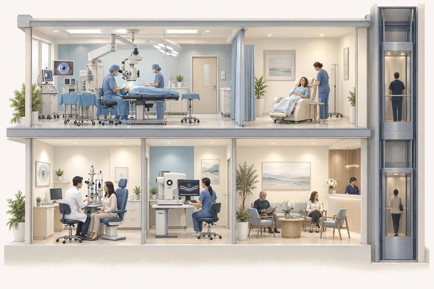
IOL exchange and complex cataractDr Chen’s clinic (level 3) and the day surgery (level 4) share one building, so assessment, surgery and follow-up happen in one place, and urgent surgery can sometimes happen very soon after assessment.
Dr Chen also operates at Chatswood Private Hospital.
Yes. All of the technology on this page is used by and available to Dr Chen at Vision Eye Institute Chatswood.
OCT stands for optical coherence tomography. It uses harmless light waves to take detailed cross-section pictures of the retina, a little like an ultrasound scan but with light instead of sound. OCT angiography (OCTA) uses the same scanner to map the retina’s small blood vessels without an injection of dye.
The Alcon UNITY Vitreoretinal Cataract System (VCS) is the surgical system Dr Chen uses for both cataract and retinal surgery. It controls pressure inside the eye in real time, and its vitreous cutter works at up to 30,000 cuts per minute.
Sometimes, for urgent problems such as certain retinal detachments. The clinic and day surgery are in the same building, but same-day surgery depends on theatre, anaesthetic and nursing availability and cannot be guaranteed.
No. Good equipment supports careful surgery, but results depend on the eye, the condition and surgical judgement. Dr Chen will explain what is realistic for your eye.
The manufacturers publish technical information for readers who want more detail. Dr Chen will explain which of these are relevant to your eye at your consultation.
For new flashes, suddenly increased floaters, a shadow or curtain, or sudden loss of vision, see the urgent assessment advice.
Clinic appointments: (02) 9424 9999