
Macular holes, pseudoholes, foveoschisis and related foveal defects: how to tell them apart on OCT
Macular holes are best distinguished by the retinal layers affected on OCT: a full-thickness hole interrupts the entire neural retina, a lamellar hole involves partial tissue loss, and foveoschisis splits the retina internally. A macular pseudohole changes the foveal contour without a true tissue defect. Tiny outer-retinal defects and atrophic cavitations can also look like holes, but have different causes and management.
For optometrists and general ophthalmologists, the useful sequence is structure first, cause second, size third. Trauma, high myopia, retinal surgery and laser injury describe why a lesion developed; “large” or “giant” describes its dimensions. These are overlapping descriptors, rather than mutually exclusive diagnoses.

How should a suspected macular hole be assessed on OCT?
Start with a high-density macular volume and closely spaced horizontal and vertical scans through the actual lesion. A single apparently normal central line can miss a microdefect, while an off-centre scan can misrepresent the opening of a full-thickness hole.
Ask three questions: is neural retinal tissue missing, is it split into layers, or is there only a steepened contour? Then assess the posterior hyaloid, any epiretinal membrane, the remaining outer retina, ellipsoid zone and RPE. Record best-corrected acuity, symptom onset, progression, refraction or axial length where relevant, and previous trauma, surgery or light exposure.
OCT and vitreomacular traction are closely linked: traction can produce a pseudocyst, schisis or a full-thickness defect. A tractional cyst with a retained outer retinal floor is not yet a full-thickness hole. Examine the fellow eye and peripheral retina as well as the macula.
| OCT finding | Working structural diagnosis |
|---|---|
| Gap through all neural retinal layers | Full-thickness macular hole |
| Irregular contour, undermined cavity and apparent tissue loss | Lamellar macular hole |
| Enclosed intraretinal split with retained roof and floor | Foveoschisis; specify the cause |
| Steep, narrow pit with foveal-sparing ERM | Macular pseudohole |
| Small focal outer-retinal defect under intact inner retina | Outer foveal defect; clarify microhole terminology |
| Cavitation or thinning in a degenerative macula | Atrophic change; determine whether a true hole coexists |
What defines a full-thickness macular hole?
A full-thickness macular hole (FTMH) is a defect extending from the internal limiting membrane through the photoreceptor layers. The RPE beneath it is not normally missing. The patient-facing macular hole page explains the condition and its treatment in simpler language.
The International Vitreomacular Traction Study classification describes FTMH by minimum linear diameter on OCT: small, ≤250 µm; medium, >250 to ≤400 µm; large, >400 µm. Measure the narrowest opening, rather than substituting the larger basal diameter. Also record whether VMT is present and whether the hole is primary or secondary to another disorder.
A stage 1 “impending hole” does not have a full-thickness defect. Size, duration, outer-retinal damage and associated disease help inform prognosis; a size label alone does not determine treatment.
How does a lamellar macular hole differ from ERM foveoschisis?
The 2020 OCT consensus separates lamellar macular hole, ERM foveoschisis and macular pseudohole. Older reports grouped some of these under “tractional” and “degenerative” lamellar holes, so historical outcome studies may mix different lesions.
For a lamellar macular hole, the mandatory findings are an irregular foveal contour, a cavity with undermined edges, and apparent loss of foveal tissue. Epiretinal proliferation, a central foveal bump and ellipsoid-zone disruption are optional features. Preserved outer retinal tissue distinguishes it from FTMH, although that outer tissue need not be entirely normal.
ERM foveoschisis requires an epiretinal membrane and schisis at the Henle fibre layer. The membrane can contract and distort the fovea, but splitting is not equivalent to tissue loss. Epiretinal proliferation associated with LMH is also not simply another name for a conventional contractile ERM.
This distinction matters when discussing surgery. Stable lesions with acceptable function may be monitored; symptomatic deterioration warrants retinal assessment. Results reported for ERM foveoschisis should not be transferred uncritically to consensus-defined LMH. See epiretinal membrane surgery or observation for functional assessment.
Is a macular pseudohole a true hole?
No. A pseudohole is usually produced by contraction of an ERM around the fovea, giving a sharply demarcated red spot and a steep, narrow pit. The consensus criteria include a foveal-sparing ERM, a steepened foveal profile and increased central retinal thickness.
There is no full-thickness tissue defect. Do not diagnose it from the fundus appearance alone or assume that every steep foveal contour is a pseudohole. OCT distinguishes this contour change from an undermined lamellar cavity. Management follows the associated membrane and its functional effect, rather than an FTMH pathway.
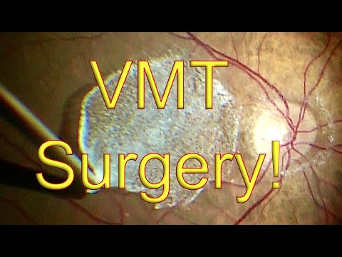
Surgery for Vitreomacular Traction by Dr. Simon Chen
 Watch on YouTube →
Dr Chen releasing vitreomacular traction during vitrectomy. This two-minute video shows real eye surgery.
What is myopic foveoschisis?
Myopic foveoschisis is splitting within the macular retina in a highly myopic eye, commonly in the setting of posterior staphyloma. It belongs to the broader spectrum of myopic traction maculopathy, which can include schisis, foveal detachment, lamellar defects, FTMH and macular-hole-associated retinal detachment.
OCT may show extensive inner or outer retinal splitting with bridging columns. Assess the posterior scleral contour, vitreoretinal traction, foveal attachment and whether an inner or outer lamellar defect is present. Schisis itself is not a full-thickness opening.
Stable schisis and a symptomatic foveal detachment have different implications; a hole with retinal detachment is different again. Surgical planning may involve vitrectomy, a macular buckle or selected combined approaches. The choice depends on traction and staphyloma configuration.
How does X-linked retinoschisis affect the macula?
X-linked retinoschisis (XLRS) is an inherited retinal disorder associated with pathogenic variants in RS1. Boys or young men may present with bilateral reduced vision and a spoke-wheel foveal appearance. OCT shows intraretinal schisis, often involving the inner nuclear layer; peripheral schisis may coexist.
The cavities are splitting within retinal tissue, not a conventional idiopathic FTMH. In older patients, schisis cavities may become less conspicuous as macular atrophy develops. Absence of a striking spoke-wheel appearance therefore does not exclude XLRS.
A bilateral pattern, age at onset, family history and peripheral findings should prompt inherited-retinal-disease assessment, with genetic testing and counselling where appropriate. Treat complications and document progression rather than applying routine macular-hole surgery to an inherited schisis phenotype.
What are macular microholes and foveal red spot syndrome?
These terms have been used inconsistently. A tiny, sharply defined foveal red spot can correspond to a focal outer-retinal defect, or occasionally a genuinely tiny full-thickness microhole. Describe the OCT depth.
The 2005 microhole series found abnormalities predominantly in the outer retina or RPE on higher-resolution OCT. The 2012 series described focal photoreceptor-region abnormalities and showed that a small lesion can affect local retinal sensitivity despite relatively good acuity. These are different from assuming that every “microhole” interrupts all neural retinal layers.
A later critical review proposed reserving “full-thickness macular microhole” for a tiny complete defect and “outer lamellar macular microhole” for the partial-thickness lesions often called foveal red spot syndrome. This is a proposed terminology refinement, not proof that older publications used the terms consistently. Nor does “outer lamellar microhole” automatically satisfy the consensus definition of LMH discussed above.
Clinically, a small central scotoma may be disproportionate to acuity loss. Dense, accurately centred OCT scans are important; the multimodal-imaging report demonstrated a subtle outer-retinal lesion that conventional imaging had not adequately characterised. Ask about light exposure and trauma, but do not attribute every red spot to either without evidence.
Many reported lesions remained stable or improved, but the evidence consists mainly of small series and case reports. Confirm the anatomy, exclude another cause of focal outer-retinal damage, and arrange follow-up according to symptoms and structural change. A tiny confirmed FTMH still needs individual retinal advice.
What does “atrophic macular hole” mean?
“Atrophic” is a descriptive qualifier, not a single standard OCT diagnosis. It may refer to a true FTMH developing in degenerating retina, an outer-retinal defect, or a cavity caused by tissue loss without a complete opening. State which structure is actually present and identify the underlying disorder.
Macular telangiectasia type 2 (MacTel2) is an important example. It can produce inner and outer retinal cavitations, an ILM drape and ellipsoid-zone loss. These degenerative cavities may occur without the retinal thickening expected from oedema; a dark OCT space is not automatically fluid or a hole. A true FTMH can develop separately.
Other relevant contexts include advanced myopic macular degeneration, longstanding inherited retinal disease and previous macular injury. Photoreceptor and RPE loss can limit visual recovery even if a true hole is closed. MacTel-associated FTMH is not invariably inoperable: surgical series report closure in selected eyes, but results from different techniques and populations should not be presented as equivalent to ordinary idiopathic holes.
How are traumatic, postoperative and laser-induced holes different?
Traumatic macular holes follow blunt ocular injury and may coexist with commotio retinae, haemorrhage, choroidal rupture or peripheral retinal damage. Establish the complete injury pattern first. Spontaneous closure is documented, particularly in selected smaller holes, so a retinal specialist may recommend a defined period of OCT observation. This is not a reason to delay the initial referral or to apply a fixed waiting period to every injury. Persistent defects need individual treatment planning.
Postoperative macular holes can occur after vitrectomy, retinal-detachment repair or membrane surgery. Direct instrument trauma is one possible iatrogenic mechanism. Delayed holes can also develop with recurrent ERM or degeneration in an already vulnerable macula. The fact that a hole follows surgery does not establish that an instrument directly injured the fovea. Compare preoperative and postoperative OCT, timing, operative findings, membrane status and retinal attachment. Published case series show that further surgery can close selected secondary holes; visual potential still depends on the original disease and residual tissue.
Laser-induced macular holes result from retinal injury rather than ordinary age-related vitreofoveal traction. High-power handheld lasers and accidental exposure to other lasers can damage photoreceptors and RPE, with or without an FTMH. Document the exposure, examine for haemorrhage or other macular injury, and confirm whether the defect is truly full thickness. Hole closure cannot reverse all associated laser damage, so anatomical and visual outcomes must be discussed separately.
Combine structure and cause: “traumatic FTMH” or “postoperative FTMH with ERM”.
What is a giant macular hole?
“Giant” describes an exceptionally large FTMH; it does not specify its cause. Definitions vary. Some recent classifications use a minimum diameter above 800 µm, while others separate a subgroup above 1,000 µm. Report the measured minimum and basal diameters and the classification used rather than implying one universal threshold.
Very large, longstanding or refractory holes may require an ILM flap or other specialist reconstructive techniques. These are selected surgical strategies, not interchangeable routine treatments. Counsel separately about closure and visual recovery, particularly where surrounding photoreceptors are damaged.
What should the referral contain, and when is it urgent?
A useful referral gives the structural diagnosis, symptom duration and change, best-corrected acuity, OCT scans and dimensions, traction or ERM, outer-retinal/RPE status, and relevant myopia, injury or surgical history. Include associated detachment and fellow-eye findings. Send actual scans where possible rather than relying only on an automated report.
Newly diagnosed FTMH warrants prompt retinal assessment. Stable LMH, pseudohole or schisis may allow planned review, but progression, diagnostic uncertainty or deteriorating function should accelerate assessment. Do not place all lesions on one urgency pathway because their names contain “hole”.
Recent ocular trauma, sudden marked visual loss, a new curtain or field defect, or symptoms suggesting retinal tear or detachment require urgent assessment. Macular-hole-associated retinal detachment also changes the urgency. Routine administration should not delay care for an acute injury or detachment. The Referrers page provides referral methods and contact details.
The central reporting rule is simple: describe what tissue is interrupted, what remains intact, and why the lesion developed. That gives the retinal surgeon more useful information than a red-spot label alone.
Where can I read more?
References
- Duker JS, et al. The International Vitreomacular Traction Study Group classification of vitreomacular adhesion, traction, and macular hole. Ophthalmology. 2013;120:2611–2619. DOI .
- Hubschman JP, et al. Optical coherence tomography-based consensus definition for lamellar macular hole. Br J Ophthalmol. 2020;104:1741–1747. Full text .
- Fung AT, et al. Epiretinal membrane: a review. Clin Exp Ophthalmol. 2021. Full text .
- Parolini B, et al. The new Myopic Traction Maculopathy Staging System. Eur J Ophthalmol. 2021;31:1299–1312. PubMed .
- Parolini B, et al. Myopic traction maculopathy: a new perspective on classification and management. Asia Pac J Ophthalmol. 2021. PubMed .
- Sieving PA, et al. X-linked congenital retinoschisis. GeneReviews. Clinical reference .
- Zambarakji HJ, et al. Macular microholes: pathogenesis and natural history. Br J Ophthalmol. 2005;89:189–193. DOI .
- Gella L, et al. Morphological and functional changes in spectral domain OCT and microperimetry in macular microhole variants. Indian J Ophthalmol. 2012;60:53–56. DOI .
- Yu S, Bellone D, Lee SE, Yannuzzi LA. Multimodal imaging in foveal red spot syndrome. Retin Cases Brief Rep. 2015;9:97–101. DOI .
- Garcia JH, et al. Macular microhole and foveal red spot syndrome: a critical review of the literature. Graefes Arch Clin Exp Ophthalmol. 2021;259:1685–1694. PubMed .
- Retinal cavitations in macular telangiectasia type 2: longitudinal structure-function correlations. Full text .
- Surgical management of full-thickness macular holes in macular telangiectasia type 2. Ophthalmology. 2024;131:66–77. DOI .
- Traumatic macular hole: clinical management and optical coherence tomography features. Full text .
- Kang HG, et al. Clinical characteristics, risk factors, and surgical outcomes of secondary macular hole after vitrectomy. Sci Rep. 2019. Full text .
- Qi Y, et al. Surgical treatment and optical coherence tomographic evaluation for accidental laser-induced full-thickness macular holes. Eye. 2017;31:1078–1084. Full text .
- Radke NV, et al. Controversy and consensus on macular hole surgery by APVRS and AAPPO. 2025. Consensus .