
Retinal vein occlusion: symptoms, treatment and what to expect
A retinal vein occlusion is a blockage in a vein that drains blood from the retina, the light-sensitive tissue at the back of your eye. It can cause bleeding and swelling inside the retina, making vision blurred or distorted. Treatment often helps the swelling and may improve sight, although recovery depends on how much the retina has been affected.

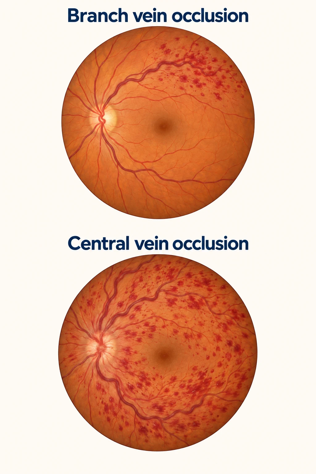
Being told that you have a “blocked vein in the eye” can be worrying. Understanding what is causing the blurred vision, and what your treatment is designed to do, can make the next steps clearer.
When does a change in vision need urgent attention?
Seek urgent medical attention straight away for sudden blurred vision or loss of sight, even if there is no pain. If an eye specialist or optometrist cannot assess you promptly, go to a hospital emergency department. Do not wait for a routine appointment or drive yourself if your vision is affected.
Several conditions can cause sudden sight loss. A retinal vein blockage cannot be diagnosed from symptoms alone, and a blockage in an artery supplying the retina is a different emergency.
What is the difference between branch and central vein occlusion?
The retina needs a steady blood supply. Arteries bring blood into it; veins carry blood away. When a vein becomes blocked, blood and fluid can leak into the surrounding retinal tissue.
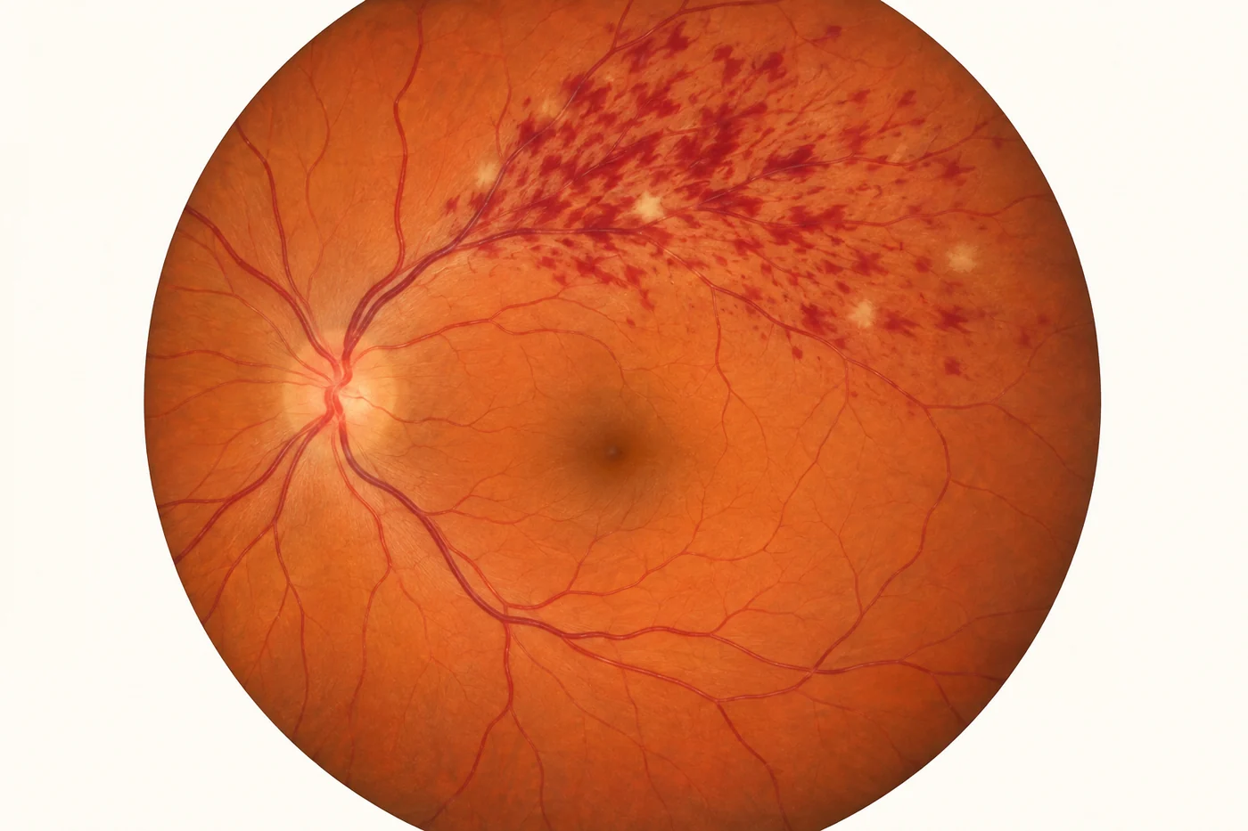
- Branch retinal vein occlusion (BRVO) affects one of the smaller veins. The changes usually involve the part of the retina drained by that vein.
- Central retinal vein occlusion (CRVO) affects the main vein draining the retina. Changes can involve a much wider area.
- Hemiretinal vein occlusion affects the drainage from roughly half of the retina.
A central occlusion can be more extensive, but the name alone does not tell you how much vision will recover. The condition of the macula and its blood supply are particularly important.
Why does a blocked vein blur my vision?
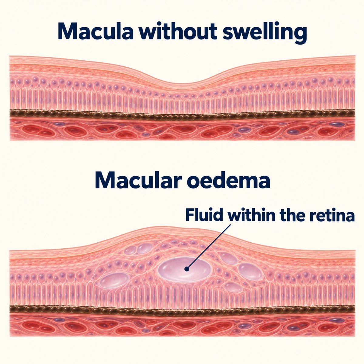
The macula is the small central part of the retina used for reading, recognising faces and seeing fine detail. Fluid leaking into it causes macular oedema, which means swelling of the macula. Words may look blurred, straight lines may appear bent, or part of the view may look dim.
Vision can also fall because the retina is not receiving enough blood and oxygen. This is called ischaemia. Swelling and poor blood supply can occur together, but they are different problems.
This distinction helps explain why a scan can show less swelling without vision returning to normal. Treatment can reduce fluid, but it cannot reliably undo damage already caused by a poor blood supply.
Why did this happen?
Retinal vein occlusion is more common as people get older. High blood pressure, raised cholesterol and glaucoma are important associated conditions. Your medical history, including diabetes and other blood-vessel problems, also matters.
Having a vein occlusion does not mean that you did something wrong. Sometimes there is no single clear explanation.
Your GP can review blood pressure, cholesterol, blood glucose and smoking, alongside your existing treatment. These checks matter for your wider health. Good control is worthwhile, but it cannot guarantee that another eye blockage will never happen.
Younger people can also develop a retinal vein occlusion. Additional investigations may be appropriate when the age, examination or medical history is unusual. A large panel of blood-clotting tests is not routinely needed for everyone.
How is retinal vein occlusion diagnosed?
Your ophthalmologist checks your vision, measures eye pressure and examines the retina, usually after drops have widened the pupil. Retinal photographs can help record the bleeding and other changes.
An OCT scan uses light to produce a detailed cross-section of the retina. It shows whether the macula is swollen and helps track its response to treatment. The scan does not involve an injection into your eye.
Sometimes an additional test is needed to assess the circulation. Fluorescein angiography follows a dye injected into a vein in your arm. OCT angiography uses a different scanning technique to show blood flow without dye. These tests answer particular questions and are not required at every appointment.
Does everyone need treatment immediately?
Not every retinal vein occlusion needs an injection. If vision is good and there is little or no swelling affecting the centre of the macula, careful observation may be appropriate. Observation still means planned checks.
When macular swelling is reducing vision, treatment is often recommended. Delaying treatment for persistent, significant swelling can limit the improvement that is possible. The decision depends on your vision, scan, blood supply and the wider condition of your eye.
How do eye injections help?
One common treatment is an injection of an anti-VEGF medicine into the eye. VEGF is a natural signal involved in blood-vessel leakage and the growth of abnormal vessels. Reducing its effect can reduce macular swelling and help vision.
The injection treats the consequences of the blockage. It does not work like a plumber removing a clot from the vein, and it is not a guaranteed cure.
Treatment often starts with injections at relatively short intervals, commonly about a month apart. The interval may later change according to your vision and OCT scan. Some people can have longer gaps or stop treatment under supervision; others need repeated treatment over several years.
Fluid can return after it has settled. A recurrence does not necessarily mean that the earlier treatment failed. It may mean that the eye still needs help controlling leakage.
A steroid treatment inside the eye is another option for selected patients. Its potential benefits need to be weighed against risks, particularly raised eye pressure and cataract. The choice is individual rather than one medicine being best for everyone.
What is having an eye injection like?
The eye is cleaned and numbed with drops before the injection. A fine needle delivers a small amount of medicine into the vitreous, the gel-filled space inside the eye. You may notice pressure or brief discomfort.
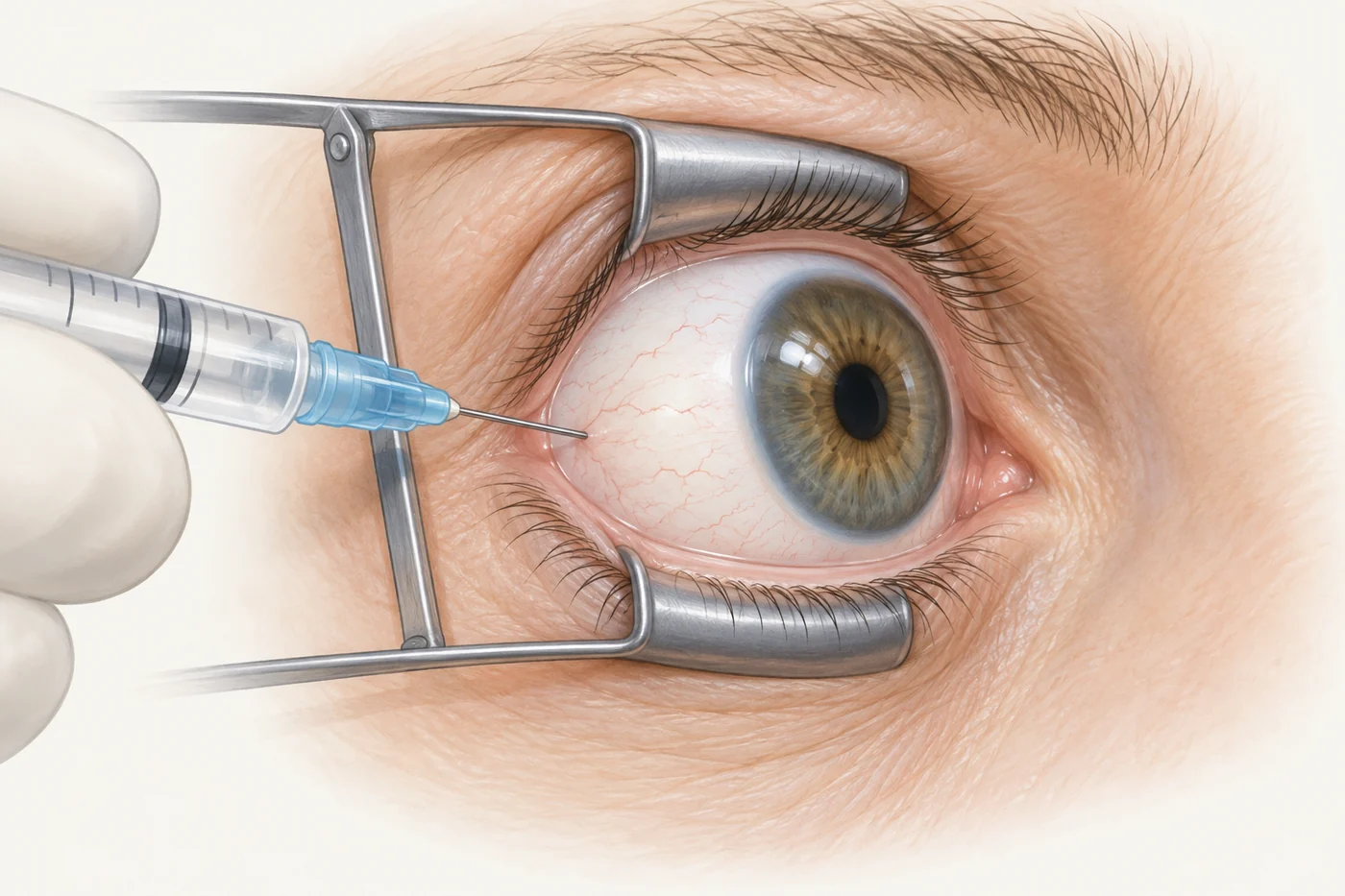
Some grittiness, a small red patch on the white of the eye or temporary blur can occur afterwards. Follow the instructions from your treating clinic rather than assuming every new symptom is normal.
Serious complications are uncommon but include infection inside the eye, severe inflammation and retinal detachment. Eye pressure can also rise. Tell the treating team about a recent stroke or heart attack, pregnancy or possible pregnancy, and any current eye infection before treatment.
Increasing pain, increasing redness or worsening vision after an injection needs urgent assessment. Contact the treating clinic immediately; if you cannot reach it, seek emergency eye care. Do not wait until the next planned injection.
When is laser treatment used?
An area of retina with a poor blood supply can stimulate fragile new blood vessels. These can bleed into the eye or grow near its drainage structures, sometimes causing a painful rise in pressure called neovascular glaucoma.
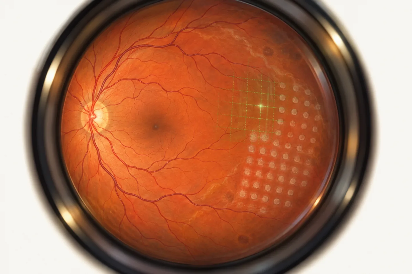
Laser may be used to treat areas of poorly supplied retina when these abnormal vessels develop. An injection may also be needed. This treatment aims to control a complication; it does not reopen the blocked vein.
In selected branch vein occlusions, laser can sometimes help manage macular swelling. It is not a routine substitute for injections in every eye, and adding laser does not reliably remove the need for further injections.
How much vision will I recover?
Some people have a substantial improvement; others retain blur or a missing area of vision despite treatment. The outlook depends on the location and extent of the blockage, how well blood is reaching the macula, the amount and duration of swelling, and the response to treatment.
Early improvement is encouraging, but one injection or one scan cannot show the whole course. Your ophthalmologist can explain what the examination and scans suggest about your own eye.
If reading, work or everyday activities remain difficult, ask about low-vision support. Better lighting, magnification and practical strategies can help you use the vision you have. You do not need to wait until treatment has finished to seek this support.
Why do I need checks when my eye feels better?
Swelling can return, and abnormal blood vessels can develop even when the eye does not initially feel different. Monitoring may therefore continue after injections are paused. It can include vision testing, OCT, eye-pressure checks and examination for new vessels.
Keep the planned appointments and contact the clinic if you cannot attend. Ask when you should next be seen and which symptoms should prompt an earlier review.
New pain, a sudden deterioration in sight, a shower of floaters or a dark curtain across the view needs urgent eye assessment. Do not assume that a new symptom is simply part of the existing vein occlusion.
Should I take aspirin or a blood thinner?
Aspirin and other blood thinners are not routinely started solely to treat a retinal vein occlusion: there is no good evidence that they reliably improve its eye outcome.
You may need these medicines for a different medical reason. Do not start, stop or change prescribed blood-thinning treatment without discussing it with the doctor responsible for it.
Can it happen again or affect my other eye?
Yes, another occlusion can occur in the same eye or the other eye, although it does not happen to everyone. Reviewing your general health and glaucoma risk is part of ongoing care. Your ophthalmologist and GP can advise which checks are relevant to you.
Before you leave an appointment, useful questions include: Is my vision affected mainly by swelling, poor blood supply, or both? What are we aiming to achieve with treatment? When is my next check? Who should I contact urgently if my symptoms change?
Sources and further reading
- Royal College of Ophthalmologists: retinal vein occlusion guideline — clinical guidance, January 2022.
- College of Optometrists: retinal vein occlusion — public explanation, updated September 2026.
- Moorfields Eye Hospital: diagnosis and treatment — patient information.
- Cambridge University Hospitals: anti-VEGF injections — the procedure and warning symptoms.
- SCORE2 trial and five-year follow-up — treatment research in central and hemiretinal vein occlusion.
- RETAIN study — longer-term follow-up in branch and central vein occlusion.
- Healthdirect: blurred vision — when to seek urgent medical attention.