
Cataract surgery after vitrectomy: what is different?
Planning the operation in an eye that has had retinal surgery
Cataract is common after vitrectomy, particularly in people over 50 who still have their natural lens. Cataract surgery can improve the blurred vision caused by the cloudy lens, but the health of the retina still matters. The operation also needs careful planning because removing the vitreous gel changes the support behind the lens.

Why might I have needed a vitrectomy?
Vitrectomy removes the clear gel from the back of the eye so the surgeon can treat a retinal problem. Common reasons include a retinal detachment, a macular hole, an epiretinal membrane that wrinkles the central retina, or blood in the vitreous that is not clearing. It is also used for selected people with persistent, disabling floaters. Many floaters do not need surgery.
Vitrectomy and cataract surgery treat different parts of the eye. Vitrectomy addresses the gel and retina; cataract surgery replaces the cloudy natural lens with a clear artificial lens. If your natural lens was already replaced before or during vitrectomy, you cannot develop another cataract in that lens.
How often does cataract develop after vitrectomy?
Cataract is a frequent longer-term effect, but there is no single percentage that applies to every eye. Studies also measure different things: the first sign of lens clouding is not the same as needing cataract surgery.
A large US registry study published in 2026 followed 181,540 eyes that retained their natural lens after vitrectomy. It estimated that 46% would have cataract surgery within two years. This describes an operation being performed, rather than every cataract that developed.
An older UK database study reported cataract surgery in about half of eyes by one year and around seven in ten by two years. These are separate studies with different patients and treatment settings, so their results should not be combined into a personal forecast.
Some cataracts become troublesome within months; others progress over several years. Age, the condition of the lens before vitrectomy, the retinal problem and whether gas or silicone oil was used can all influence the course.
Why does removing the vitreous affect the lens?
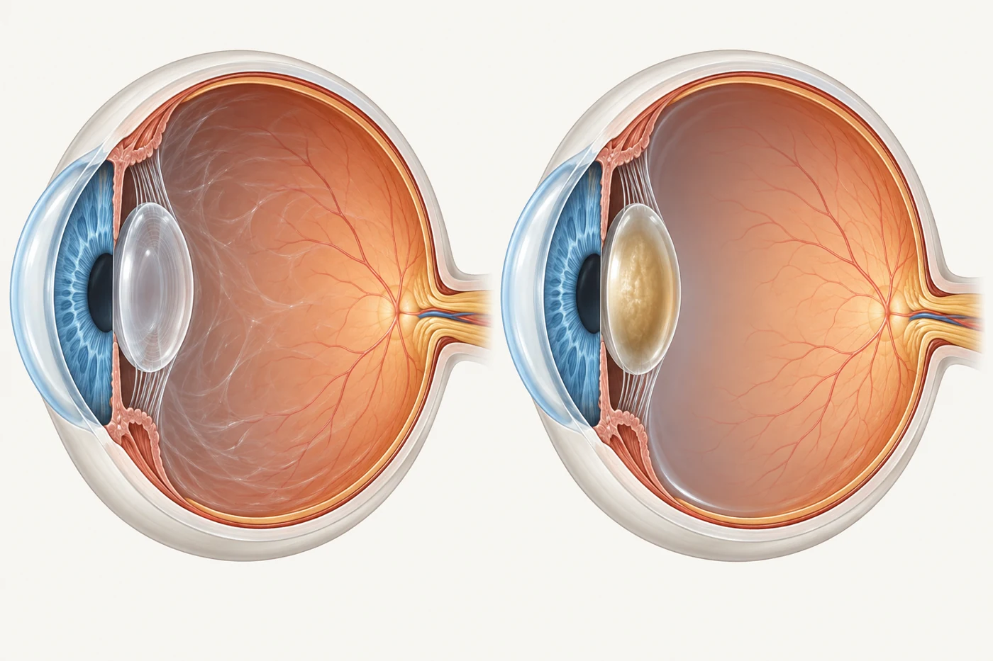
The vitreous helps maintain the environment around the natural lens. After it is removed, the lens is exposed to more oxygen. This is thought to contribute to changes in lens proteins and the development of a nuclear cataract—a cloudy, often yellowing centre of the lens. Cataract can therefore develop after an otherwise uncomplicated vitrectomy; its appearance does not, by itself, mean the lens was damaged during the operation.
The eye is not left empty. Fluid fills the space previously occupied by the gel. A gas bubble may be used temporarily, or silicone oil may be left inside for retinal support, with a separate plan for its management.
Removing the vitreous gel changes the environment behind the lens. The right-hand image shows a possible later cataract, not an inevitable or immediate result. Illustration only; the eye remains filled.
Does cataract develop faster in older people?
Usually, yes. A study of 301 eyes operated on for macular hole or epiretinal membrane found that nuclear cataract progressed much more slowly in people under 50 than in those over 50. However, progression was similar across the older age groups: each extra decade did not bring a predictable increase.
Fifty is not a biological cut-off. A younger person may keep a clear lens for years, but can still develop a cataract, including clouding near the back of the lens. A small study specifically examining patients under 50 confirmed that cataract changes can occur in this age group.
Will cataract surgery restore my vision?
It can improve the part of your visual difficulty caused by the cataract. It cannot reverse damage to the retina. For example, distortion from an epiretinal membrane or reduced central vision after a retinal detachment may remain even after the lens is clear. Large studies show useful improvement after postvitrectomy cataract surgery, but average final vision is lower than in eyes without a history of vitrectomy.
Before recommending surgery, the surgeon assesses how much of the blur comes from the lens and how much comes from the retina. Examination and an OCT scan of the macula—the central retina—help with that discussion. They improve planning but cannot predict the final result with certainty.
What makes the cataract operation different?
The same basic operation is used: the cloudy lens is removed through a small incision and replaced with an intraocular lens, or IOL. The difference is how the tissues behave during surgery.
Can the zonules be weaker?
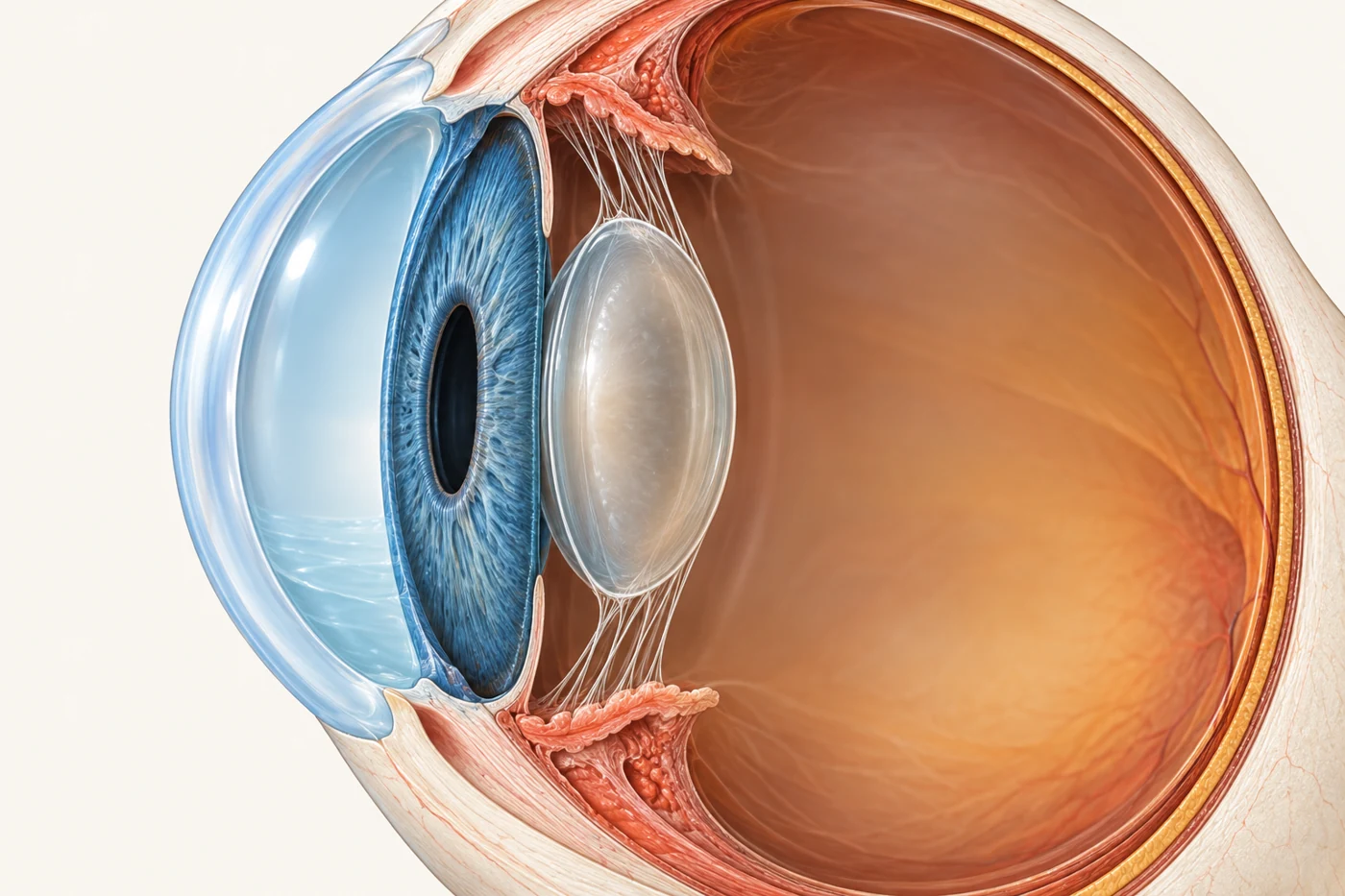
Zonules are fine fibres that suspend the lens and its thin surrounding capsule. They can be weaker in some previously vitrectomised eyes. If support is poor, the surgeon may need additional support for the capsule or another way to secure the implant.
What changes when there is no vitreous gel support?
Fluid remains behind the lens, but the gel's mechanical support is lost. The capsule may move more during surgery and needs careful handling.
Why can the anterior chamber become deeper?
The anterior chamber is the space between the cornea and iris at the front of the eye. During cataract surgery, irrigation fluid can push the iris and lens backwards more readily after vitrectomy. The chamber may become unusually deep or change depth as the operation proceeds, particularly in long, short-sighted eyes. Careful fluid control and gentle handling help manage these changes.
Fine zonular fibres hold the lens capsule in position. After vitrectomy, the surgeon assesses both these fibres and the changed support behind the lens. This static illustration does not show a torn zonule or predict how an individual eye will behave during surgery.
Are complications more likely?
Some complications are more frequent, but the increase is not the same for every complication. A UK study compared 2,221 cataract operations after vitrectomy with 136,533 operations in other eyes. Zonular damage during surgery occurred in 1.3% versus 0.6%, and lens fragments dropped into the back of the eye in 0.6% versus 0.2%. Posterior capsule rupture—a tear in the back of the lens capsule—was similar: 1.5% versus 1.7%.
These figures describe study groups, not your individual risk. The reason for the original vitrectomy, the state of the capsule and zonules, and any other eye conditions all need to be considered.
Why does the surgeon's experience matter?
Relevant experience helps the surgeon anticipate these anatomical changes and plan for unexpected difficulties, with access to retinal expertise if needed.
Useful questions are whether the surgeon regularly manages eyes like yours, how the retinal condition affects the expected result, and what the plan would be if the capsule cannot support the intended implant. No level of experience removes every risk.
Is choosing the lens implant more difficult?
Lens measurements and the glasses prescription being targeted deserve careful attention. Silicone oil, a long eye or previous retinal-detachment surgery using a scleral buckle can affect lens-power planning. A study of 974 previously vitrectomised eyes found that the accuracy of different calculation methods varied with these features. Glasses may still be needed after surgery.
Macular disease may make multifocal lenses less suitable. A toric implant may still help suitable corneal astigmatism. Lens design needs an individual plan. Read more about lens implant choices.
When should cataract surgery be considered?
An early cataract can be monitored. Consider surgery when it impairs useful vision or retinal assessment, coordinating timing with retinal recovery and any gas or oil. There is no universal waiting period.
If vitrectomy has not yet taken place, it may be worth discussing whether cataract and retinal surgery should be combined. For a later cataract operation, bring details of your previous retinal surgery and discuss both the expected visual benefit and the additional surgical considerations.
What needs attention during recovery?
Follow-up checks both the front and back of the eye. Inflammation, eye-pressure changes or swelling at the macula can affect recovery and may need treatment. Persistent blur should be assessed rather than assumed to be part of normal healing.
New flashing lights, a sudden increase in floaters, a curtain or shadow in your vision, or sudden visual loss need urgent eye assessment. Contact your surgeon's urgent-care service or an emergency eye service rather than waiting for a routine appointment.
For a planned assessment with Dr Chen, arrange an appointment through Vision Eye Institute Chatswood. The vitrectomy and cataract surgery pages explain these operations in more detail.
References and further reading
- Wang JC et al. Cataract Progression and Risk Factors for Cataract Surgery after Pars Plana Vitrectomy in Phakic Eyes: An IRIS Registry Analysis. Ophthalmology Retina. 2026.
- Jackson TL et al. United Kingdom National Ophthalmology Database Study of Vitreoretinal Surgery: report 1. Eye. 2013.
- Thompson JT. The role of patient age and intraocular gas use in cataract progression after vitrectomy for macular holes and epiretinal membranes. American Journal of Ophthalmology. 2004.
- Kataria AS, Thompson JT. Cataract Formation and Progression in Patients Less Than 50 Years of Age after Vitrectomy. Ophthalmology Retina. 2017.
- Soliman MK et al. A Database Study of Visual Outcomes and Intraoperative Complications of Postvitrectomy Cataract Surgery. Ophthalmology. 2018.
- Markatia Z et al. The Post Vitrectomy Cataract. International Ophthalmology Clinics. 2022.
- Rey A et al. Visual outcome and complications of cataract extraction after pars plana vitrectomy. Clinical Ophthalmology. 2018.
- Zhang J et al. Choice of intraocular lens calculation formula for cataract patients with prior pars plana vitrectomy. Journal of Cataract and Refractive Surgery. 2023.
- Holekamp NM, Shui YB, Beebe DC. Vitrectomy surgery increases oxygen exposure to the lens: a possible mechanism for nuclear cataract formation. American Journal of Ophthalmology. 2005.
- ASRS: vitrectomy
- vitrectomy for floaters
- Wang and colleagues
- Thompson
- Rey and colleagues
- Holekamp and colleagues
- Kataria and Thompson
- Soliman and colleagues
- Markatia and colleagues
- Zhang and colleagues
- ASRS: retinal detachment
These are the published sources behind this article. They are written for clinicians and do not replace advice about your own eye.
How do I make an appointment with Dr Chen?
Appointments and enquiries through Vision Eye Institute, Chatswood.
Clinic appointments: (02) 9424 9999