
Cataract surgery with myopia: retinal risks and lens choices
Cataract surgery can improve vision and reduce short-sightedness, but a highly myopic eye needs extra planning. The main decisions are how to protect and assess the retina, which lens implant to use, and whether to aim for distance vision, preserve some near vision or use monovision.

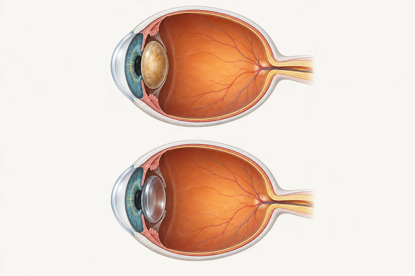
If you have spent years taking your glasses off to read, that habit matters. A clear distance result can be welcome, but losing familiar close vision can be a surprise unless it is discussed before surgery. Equally, removing a cataract cannot repair damage already present in the retina.
This guide explains the choices for patients. A technical section at the end covers additional points for referring optometrists.
Why does short-sightedness change the planning?
In many people with myopia, the eye is longer than usual from front to back. This is called
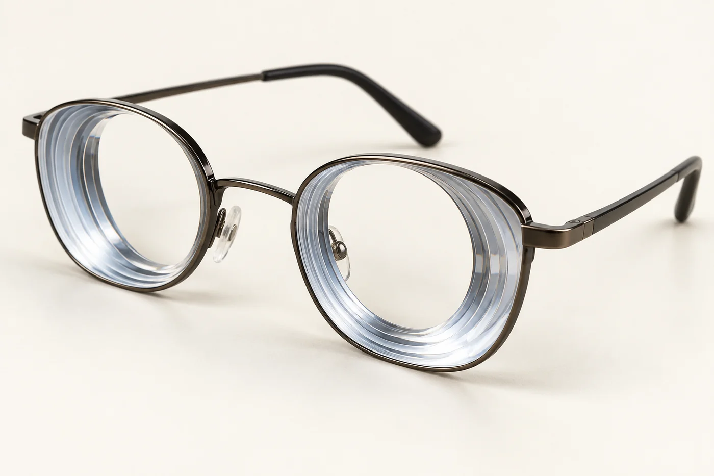
axial myopia. A longer eye changes the lens power needed to focus light and may also have thinner or more vulnerable retinal tissue.
Your glasses prescription is only part of the picture. Cataract itself can cause an increasing minus prescription, and previous laser vision correction can hide how short-sighted you once were. The surgeon therefore considers the eye's measured length, its retinal health and its history, rather than relying on the current glasses prescription alone.
High myopia and myopic retinal disease are not interchangeable. Some very short-sighted people have healthy central retinas. Others have stretching, atrophy, abnormal blood vessels or traction affecting the macula—the part used for reading and recognising faces. These changes may limit the improvement possible after cataract surgery.
How much does the risk of retinal detachment increase?
Retinal detachment occurs when the light-sensitive lining at the back of the eye separates from the tissue beneath it. A tear can allow fluid under the retina. Short-sighted eyes are already more susceptible, and cataract surgery adds to that risk.
The risk is not the same for everyone. It is influenced by age, eye length, sex, retinal findings, previous retinal problems and whether surgery is complicated by damage to the lens capsule or loss of vitreous gel. A previous detachment in either eye is particularly relevant to the discussion.
Two studies show why a single percentage can be misleading:
- Across a broad US cataract population, the IRIS Registry recorded retinal detachment in about 2 in 1,000 operated eyes within one year. This included people aged 40 and over and was not a study solely of high myopia.
- In a selected higher-risk Swedish subgroup—men younger than 60 with an eye length of at least 25 mm—about 9 in 100 eyes developed detachment during follow-up averaging roughly 4.7 years in the overall study. This is not the expected risk for every short-sighted patient.
These figures have different populations and observation periods. They should not be compared as if surgery increased everyone's risk from the first figure to the second. Your surgeon can explain which features apply to your eyes and the uncertainty in any estimate.
Risk continues beyond the early recovery period. A good result at the first postoperative visits does not mean retinal symptoms can be ignored years later.
What retinal checks are needed before surgery?
A careful dilated examination should assess the peripheral retina as well as the macula. The far edge of the retina is where important tears and areas of lattice degeneration may lie. Lattice is a pattern of peripheral retinal thinning; it is not itself a retinal detachment.
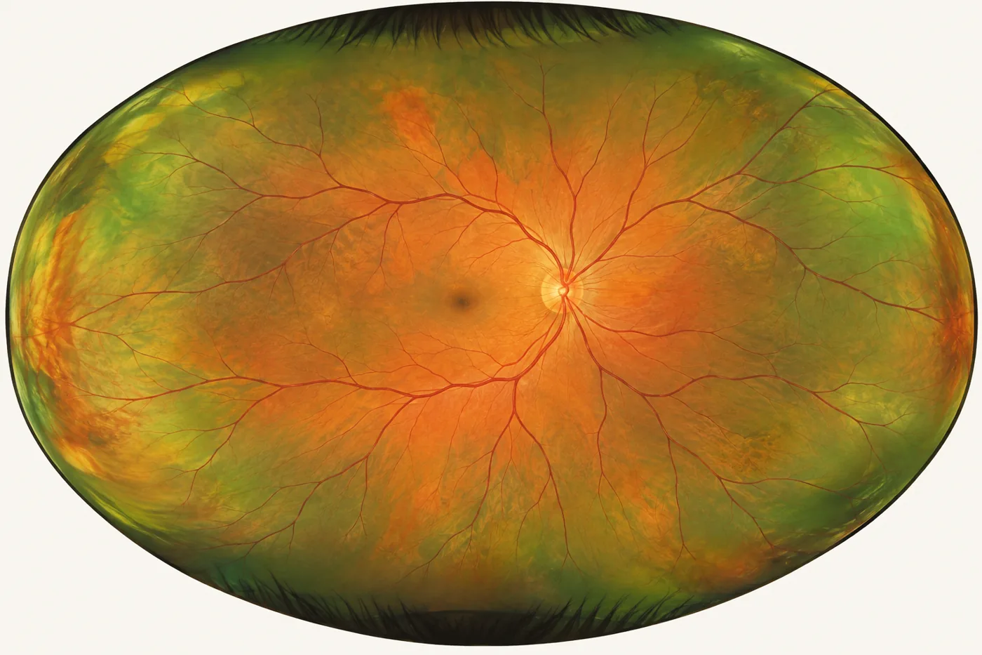
The examination may include gentle pressure on the outside of the eye, called scleral depression, to bring the far periphery into view. Widefield photographs help document findings, but do not replace a careful examination. An OCT scan provides detailed information about the macula; it does not clear the peripheral retina of tears.
If the cataract is too dense to see through, an ultrasound scan can look for a detachment or other major abnormality. It cannot reliably exclude every small tear. The plan should include examining the retina again when the view becomes clearer after surgery.
Tell the surgeon about previous retinal laser, a detachment or vitrectomy, recent flashes or floaters, and retinal detachment in close relatives. Bring earlier reports where available.
Should retinal tears or lattice be lasered first?
A significant retinal tear may need treatment before cataract surgery. Not every peripheral retinal finding needs preventive laser.
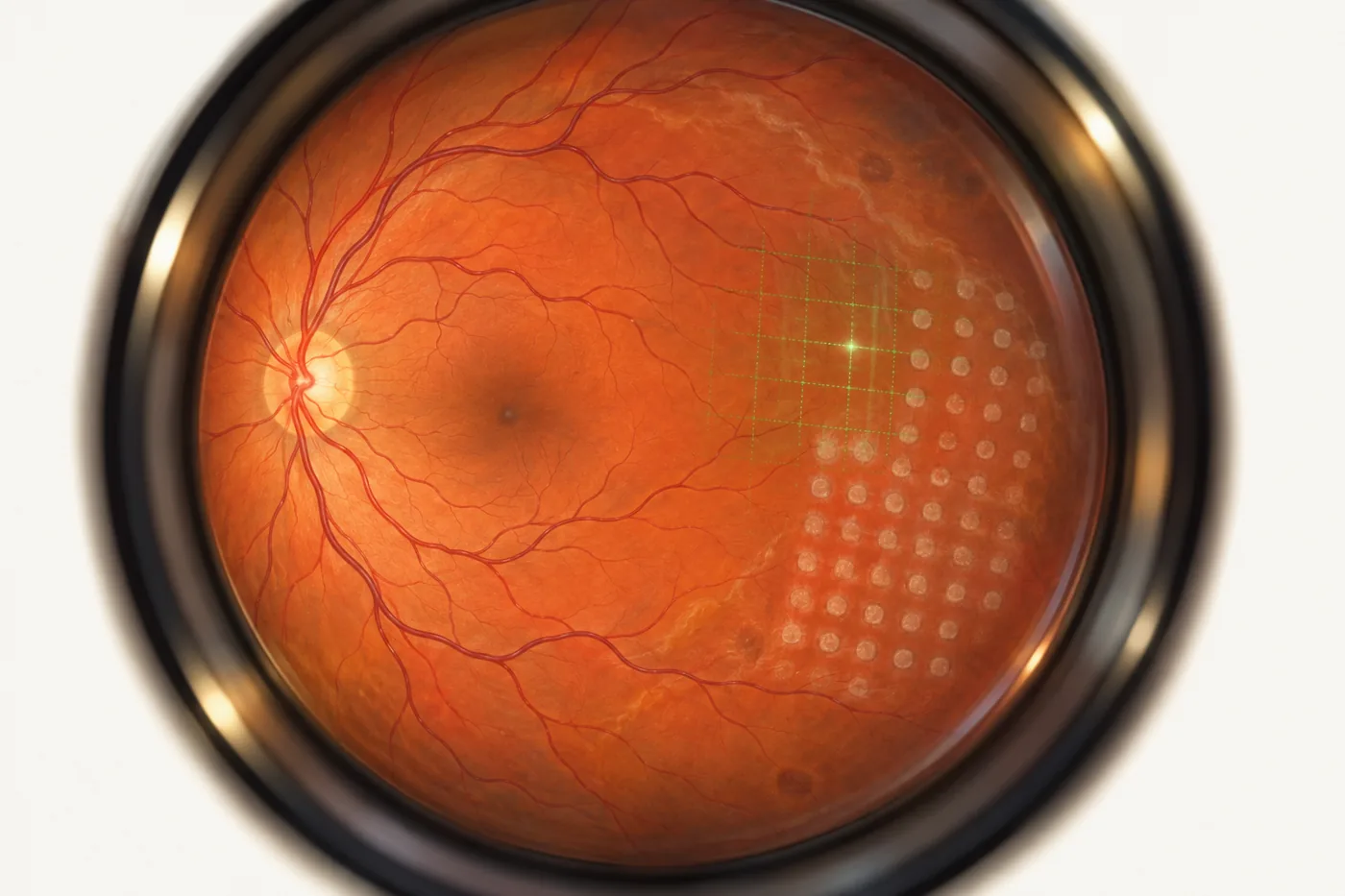
A fresh horseshoe-shaped tear with symptoms and ongoing pulling on the retina usually needs prompt treatment. Laser, or occasionally freezing treatment, creates a surrounding adhesion to reduce the chance of fluid passing beneath the retina. If a detachment has already developed, retinal surgery may be needed instead.
An asymptomatic tear is assessed differently from a small atrophic hole or lattice. Lattice without a tractional tear, and many small holes without progressive fluid, can be observed. Current guidance finds insufficient evidence to recommend routine preventive treatment of all asymptomatic breaks simply because cataract surgery is planned.
There are grey areas, including an eye with lattice when the other eye has already detached. Lesion type, surrounding fluid, symptoms, vitreous changes and the wider history guide an individual decision. An absence of strong trial evidence does not mean treatment can never be appropriate; it means the benefits and harms need a specific reasoned discussion.
After treatment, the retinal specialist decides when the retina is sufficiently stable to proceed. Laser reduces risk from the treated area but does not prevent every future tear elsewhere.
Should I aim for distance vision or stay a little short-sighted?
The target refraction is the glasses prescription the surgeon aims to leave after healing. It is a target, not a guarantee. There are three useful approaches.
Distance vision in both eyes
Aiming close to zero prescription—called emmetropia—can make distance activities easier without glasses. With standard monofocal implants, reading glasses will usually still be needed.
This can feel very different if you have always read by taking your distance glasses off. A lens aimed for distance does not recover the natural lens's youthful ability to change focus.
Some short-sightedness in both eyes
Keeping a planned amount of myopia may preserve useful unaided vision for reading or other close work. Distance glasses are then expected. The amount should reflect your preferred working distance, rather than automatically reproducing a very strong preoperative prescription.
For example, someone who values reading in bed without glasses may make a different choice from someone whose priority is distance vision for outdoor activities. The surgeon and optometrist can help translate that preference into a realistic target.
Monovision or a smaller difference between the eyes
With monovision, one eye is aimed mainly for distance and the other for nearer tasks. A smaller difference, often called mini-monovision, may favour distance and computer-range vision. It generally provides less unaided fine reading than a stronger near target.
Some people adapt comfortably; others dislike the imbalance. Depth perception and the quality of binocular vision can be affected, and glasses may still help for driving, low light or prolonged reading. Previous successful contact-lens monovision is useful information. A contact-lens trial may help when the cataract still allows a meaningful test.
Whichever approach you prefer, both eyes need a coordinated plan. A large temporary prescription difference after first-eye surgery can make ordinary glasses difficult to tolerate. Discuss the second eye's vision, timing and interim correction before choosing the first eye's target.
Which type of lens implant makes sense in a myopic eye?
There is no single best implant for all short-sighted patients. Retinal health, desired focus, astigmatism, night-vision needs and the available lens powers all matter.
- Monofocal lenses provide one main focus. They are often a practical choice when retinal disease makes preserving image quality the priority. The focus can be aimed for distance or near.
- Toric lenses also correct regular corneal astigmatism. A toric lens is not necessarily multifocal: it can have a single focus. Accurate measurements and rotational stability matter.
- Enhanced monofocal or extended-depth-of-focus lenses may broaden the useful range of vision. The benefit and optical compromises vary by model; reading glasses may still be needed.
- Multifocal or trifocal lenses offer several useful focal distances but can produce haloes, glare and reduced contrast. Existing macular disease, or concern about future retinal function, can make those compromises less acceptable.
Being myopic does not automatically rule out every lens that extends the range of vision. However, evidence from otherwise healthy eyes should not be assumed to apply to an eye with myopic macular damage. Some models are also unavailable in the very low powers a long eye needs.
You can read more about the general categories in the.
Why might I need a low-power, zero-power or negative-power lens?
An exceptionally long eye may need very little additional focusing power after its cataract is removed. The calculation may therefore call for a low positive-power implant, a zero-power implant, or a negative-power implant.
The number printed on the implant is not your eventual glasses prescription. A negative-power IOL can be the correct choice for a long eye being aimed at good distance focus. Conversely, an implant with positive power can be used while deliberately leaving a person short-sighted.
Specialist ranges make these cases possible, but the combination of power, astigmatism correction, material and design may be narrower than for an average-length eye.
What is different about ZEISS AT TORBI?
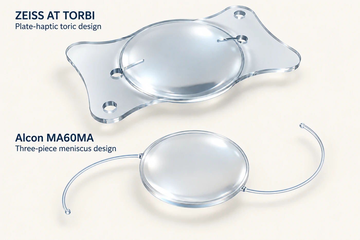
AT TORBI is ZEISS's toric lens family. Its plate-shaped supports look quite different from the thin curved arms of a three-piece lens. It corrects astigmatism as well as providing the required focusing power.
For example, the international AT TORBI 709M datasheet lists sphere from −10 to +32 D, with cylinder from +1 to +12 D. That makes it relevant when a very low power and substantial astigmatism correction are both needed. These are implant specifications, not spectacle powers.
Model numbers matter. The 719M uses spherical-equivalent labelling, and preloaded MP versions can have different ranges. The exact model and orderable sphere/cylinder combination must be checked rather than assuming the entire family is interchangeable.
These lenses are hydrophilic acrylic with a hydrophobic surface. That material distinction is relevant when considering possible future retinal surgery, as discussed below.
What is an Alcon negative-power meniscus lens?
The Alcon AcrySof MA60MA is a three-piece lens: an acrylic optic with two separate fine PMMA supporting arms, called haptics. Model-specific documentation gives a range of −5 to +5 D in 1 D steps. It has a 6 mm optic and a 13 mm overall length.
Its meniscus optic has a concave surface and a convex surface, rather than the familiar two outward-curving surfaces of a conventional biconvex lens. A meniscus design can provide low or negative power; its shape alone does not tell you the precise power. The MA60MA is not a toric lens, so corneal astigmatism needs a separate plan.
The related Alcon MA60AC is a different model. Its specifications should not be substituted for those of the MA60MA just because the names look similar.
Are there other extended-range or custom options?
Yes. For example, the Australian listing for the non-toric ZEISS CT ASPHINA 404 extends down to −10 D. It offers a different combination of focusing power, support design and material from the Alcon MA60MA. It is also hydrophilic acrylic with a hydrophobic surface.
Some unusual prescriptions require a special order or consideration of custom manufacture. An extended-range catalogue lens is not necessarily a bespoke lens made uniquely for one person. The surgeon needs to confirm the exact power, design, local supply arrangements and lead time before finalising surgery. Australian listing does not mean every power is sitting on a local shelf.
Does lens material matter if I might need retinal surgery later?
Yes. The implant should be considered alongside possible future retinal treatment, particularly if the eye already has significant retinal disease.
Some complex detachments need silicone oil inside the eye to support the retina. Oil can adhere firmly to a silicone lens implant, obscuring the view and affecting vision. This is a reason to consider avoiding silicone IOLs when future silicone-oil treatment is a realistic concern. It does not mean that silicone IOLs themselves cause retinal detachment.
There is a different issue with hydrophilic acrylic lenses: calcium deposits and clouding have been reported after retinal surgery involving gas. A hydrophobic surface does not make a hydrophilic lens identical to a lens made from hydrophobic acrylic throughout.
For these reasons, a suitable hydrophobic acrylic lens may be preferred when future retinal intervention is a significant possibility. It is not immune to all deposits or complications. When the required power or cylinder limits the available options, the surgeon must balance retinal considerations against the optical needs of that particular eye.
What can make the operation technically different?
A long eye can have a deeper front chamber, more flexible supporting tissues and a larger lens capsule. The surgeon may need to adjust the operating fluid flow, instrument position and the way the lens capsule is supported.
One recognisable problem is reverse pupillary block, also called lens–iris diaphragm retropulsion syndrome. During surgery, fluid pressure can press the pupil edge against the front of the lens capsule, temporarily creating a seal. The iris and lens move backwards, the front chamber becomes unusually deep and the pupil may enlarge. This can cause discomfort.
Recognising the pressure imbalance allows the surgeon to restore communication between the fluid compartments and adjust the surgical conditions. Previous vitrectomy can make this behaviour more likely. It is an intraoperative issue for the surgical team to manage, not something a patient needs to prevent.
Other considerations include protecting weak lens-supporting fibres, keeping the chamber stable, ensuring the wound seals and achieving secure implant positioning. A toric lens must remain at its intended angle. Extra capsular support may be useful in selected eyes, but is not required solely because someone is short-sighted.
Does myopia affect the choice of anaesthetic?
It can. A very long eye, particularly one with a posterior outpouching called a staphyloma, changes the anatomy around the eye. A sharp-needle injection beside or behind the eye carries a rare but serious risk of penetrating or perforating the globe.
Options can include numbing drops, anaesthetic placed inside the front chamber during surgery, or a sub-Tenon block delivered using a blunt cannula. These techniques have different benefits and limitations. Avoiding a sharp orbital needle avoids that particular needle mechanism of injury; it does not make the whole procedure risk-free.
The surgeon and anaesthetist consider eye shape, the planned operation, comfort and the ability to remain still. Sedation or general anaesthesia may be appropriate in selected circumstances. High myopia alone does not mean that general anaesthesia is necessary.
How accurately can the final prescription be predicted?
Modern measurements and formulas have improved lens selection, but very long eyes remain more difficult to predict. The surgeon measures the eye's length and corneal curvature, checks the quality of those measurements and calculates the likely result with the intended implant.
An irregularly stretched back wall, poor fixation, a dense cataract or previous corneal laser surgery can complicate the calculations. Comparing suitable modern formulas helps identify unexpected results, but agreement between formulas cannot rescue an incorrect measurement.
A result that is more long-sighted than intended can be particularly disappointing for someone used to easy unaided reading. The surgeon may discuss a modest myopic target in some circumstances, but it should reflect your priorities and the measured uncertainty—not a standard rule for every myopic eye. Glasses, contact lenses or occasionally further treatment may still be needed.
Which symptoms need urgent attention after surgery?
New flashes, a sudden shower or increase of floaters, a curtain or shadow in the vision, or sudden loss of sight need urgent same-day assessment. Contact the operating team or an emergency eye service. If you cannot reach them promptly, attend an emergency department. Do not wait for the next routine appointment, even if you have had retinal laser before.
Increasing pain or worsening vision after cataract surgery also needs urgent advice, because retinal detachment is only one possible cause. Keep the emergency contact instructions supplied by your surgical team. Longer-term eye examinations remain important after the cataract has healed.
What should referring optometrists include and consider?
The following section adds technical detail to the patient discussion above.
Referral information that changes the plan
Include historical and current refraction, best-corrected acuity, axial length if available, recent refractive change, and the extent to which symptoms fit the cataract. Document previous LASIK, PRK or radial keratotomy; a small current prescription does not exclude a long eye. Prior retinal tears, laser, detachment, vitrectomy or scleral buckling—and the status of the fellow eye—are particularly useful. Include available macular OCT and peripheral findings without presenting an image as a substitute for an examination.
Record the patient's habitual reading distance, whether they remove glasses to read, previous monovision tolerance, binocular problems and the preferred postoperative target. Clarify if a temporary anisometropia is likely to be difficult. These details can be as important as the measured spherical equivalent. Use the existing for routine referrals; new retinal symptoms warrant urgent clinical assessment.
How should retinal-risk figures be interpreted?
The IRIS one-year RRD result was 6,690/3,177,195 eyes (0.21%). In the Swedish study,
298/58,624 eyes (0.51%) detached over mean follow-up 4.67 years. The under-60 male, AL ≥25 mm subgroup was 44/465 (9.46%). Keep these denominators and time horizons intact. The 25 mm study threshold is not a universal definition of high myopia.
Neither registry isolates the causal effect of surgery from all underlying myopic risk. Coded lattice and other associations in IRIS should not be converted from odds ratios into an individual absolute risk or multiplied together. The 2026 Babu cohort adds longer-term evidence but uses patients after bilateral phacoemulsification; its outcomes are not interchangeable with eye-level rates. A normal peripheral examination or prior retinopexy does not confer lifetime clearance.
What deserves extra attention in biometry?
Check measurement repeatability, fixation and the actual axial-length trace or scan, particularly with posterior staphyloma or discrepant fellow-eye values. The relevant distance is to the functioning foveal region; an ultrasound measurement towards the deepest part of an eccentric staphyloma can be misleading. Optical biometry is generally preferred when a reliable reading can be obtained; dense media may require ultrasound with appropriate attention to alignment.
Optimise the ocular surface and investigate inconsistent corneal readings. For toric planning, assess regularity, posterior corneal astigmatism and the model-specific cylinder convention. Previous LASIK/PRK/RK requires a post-refractive-surgery calculation pathway, not an ordinary long-eye formula used without modification. Prior buckling, current silicone oil and other altered anatomy also need explicit consideration.
What are the strengths and limitations of the formulas?
Barrett Universal II, Kane, EVO and Olsen: modern options with encouraging results in extreme axial-length studies. In Guo's 73-eye AL ≥29 mm series, Kane, EVO 2.0, Barrett Universal II and Olsen had similar median absolute errors, approximately 0.29–0.34 D, compared with
0.80–0.85 D for Haigis and SRK/T. Kane's signed mean error was closest to zero. This was a single-model Rayner 920H dataset, so it does not prove the same ranking for every low or negative IOL.
Hill-RBF: a data-driven alternative. In the 35-eye AL >28 mm series, Hill-RBF 3.0 was broadly comparable with Kane, Barrett Universal II and EVO. The version and validated measurement range matter, especially near an extreme of the training data.
SRK/T and Holladay 1 with axial-length adjustments: Wang–Koch or Cooke-modified axial length can improve selected older-formula predictions. The 35-eye series found SRK/T with CMAL competitive with the modern formulas, while Holladay 1 with either adjustment performed less well. An adjustment is formula- and implementation-specific; it should not be added indiscriminately to a modern calculator or to measurements already corrected by another method.
Haigis: remains a useful established approach, using measured anterior chamber depth and lens constants. Its performance in moderately long eyes should not be assumed to hold at very extreme lengths; it performed less well in the cited ≥29 mm series. That result does not make it universally unsuitable.
Cross-check plausible outputs using constants appropriate to the exact lens, biometer and formula. Investigate discrepancies rather than averaging incompatible outputs. Pay attention to low/zero/negative-power models and the available power steps. Specify the intended refractive target separately from any correction intended to address systematic formula bias. No formula can promise an exact result or overcome macular pathology.
Which implant and surgical details merit explicit handover?
Distinguish optic power from target refraction, sphere from spherical equivalent, and IOL-plane cylinder from spectacle cylinder. The 709M international datasheet uses sphere; Australian 719M listings use spherical equivalent. Do not apply the broad range of the grouped Alcon AL004 billing entry to MA60MA alone. Confirm the actual model, ordered power, packaging and current instructions with the surgical service.
Document the rationale if a hydrophilic extended-range lens is selected in an eye that may need future gas tamponade, or if silicone material is being considered where future silicone oil is plausible. Neither concern supplies a reliable numerical complication probability. Toric rotational stability, zonular status, capsular support and the consequences of a refractive surprise also belong in the discussion.
Flag previous vitrectomy, deep chamber anatomy and staphyloma to the surgical/anaesthetic team. The postoperative plan should distinguish expected refractive rehabilitation from symptoms requiring urgent examination and from continuing surveillance of myopic retinal disease.
Sources and further reading
The sources below support the retinal, refractive, implant and anaesthetic discussions. Device specifications and availability should be checked again when an individual lens is ordered.
- AAO retinal examination and treatment recommendations, December 2024 summary benchmark .
- Morano and colleagues: retinal detachment and tears after cataract surgery in the IRIS Registry, 2023 .
- Thylefors and colleagues: Swedish population study of retinal detachment after cataract surgery, 2022 .
- Babu and colleagues: retinal detachment and tears up to ten years after phacoemulsification, 2026 .
- ESCRS recommendations for cataract surgery .
- Lens–iris diaphragm retropulsion syndrome: incidence and risk factors, 2017 .
- ESCRS: technical considerations in phacoemulsification in high myopia .
- Guo and colleagues: formula accuracy in eyes with axial length at least 29 mm, 2022 .
- Moshirfar and colleagues: six formulas in eyes longer than 28 mm, 2022 .
- ZEISS AT TORBI 709M and 709MP technical datasheet .
- ZEISS toric IOL family .
- Alcon MA60MA model specifications: PMDA-hosted instructions in Japanese .
- Alcon historical catalogue: multipiece and meniscus IOLs, printed page 50 .
- Australian Government Prescribed List, July 2026 XML data .
- Apple and colleagues: silicone oil adhesion to silicone IOLs, 1996 30463-6).
- Silicone oil interactions with other IOL materials, 2001 .
- Bopp and colleagues: IOL calcification after vitrectomy, 2023 .
- Globe penetration during local anaesthesia: clinical series, 2022 .
- NICE cataract guidance: anaesthesia and preoperative assessment .