
Alcon UNITY VCS: cataract and retinal surgery technology explained
Alcon UNITY VCS is the machine that powers and controls several of the instruments used in cataract and retinal surgery. It helps the surgeon remove a cataract or vitreous gel, balance fluid moving through the eye, and manage surgical lighting and compatible retinal laser instruments.

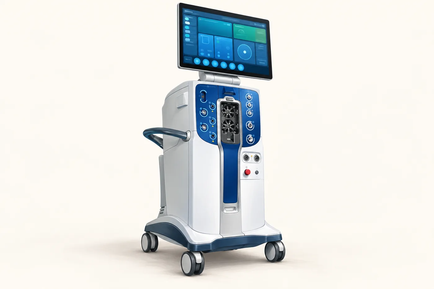
Its main developments concern control: how ultrasound breaks up a cataract, how the eye is supported as fluid leaves it, and how the system responds to changes in suction or heat. Understanding these jobs makes names such as
4D Phaco, Intelligent Sentry and Light Sentry much less mysterious.
What is UNITY VCS, and where does it fit in the operation?
VCS stands for Vitreoretinal Cataract System. “Vitreoretinal” refers to the vitreous gel inside the eye and the retina lining its back wall. The platform supports both anterior-segment surgery, including cataract removal, and posterior-segment surgery, including vitrectomy. Alcon also makes UNITY CS, a cataract-only version.
The console stays beside the operating table. Tubing and cables connect it to small hand-held instruments. The surgeon looks through a microscope or a surgical viewing system, directs the instruments and uses a foot controller to vary their action. The console supplies and regulates functions in response.
It does not choose where to operate, select a lens implant or perform the operation independently. A machine's capabilities also depend on its installed modules, compatible instruments and the settings chosen for that eye.
What does 4D Phaco mean?
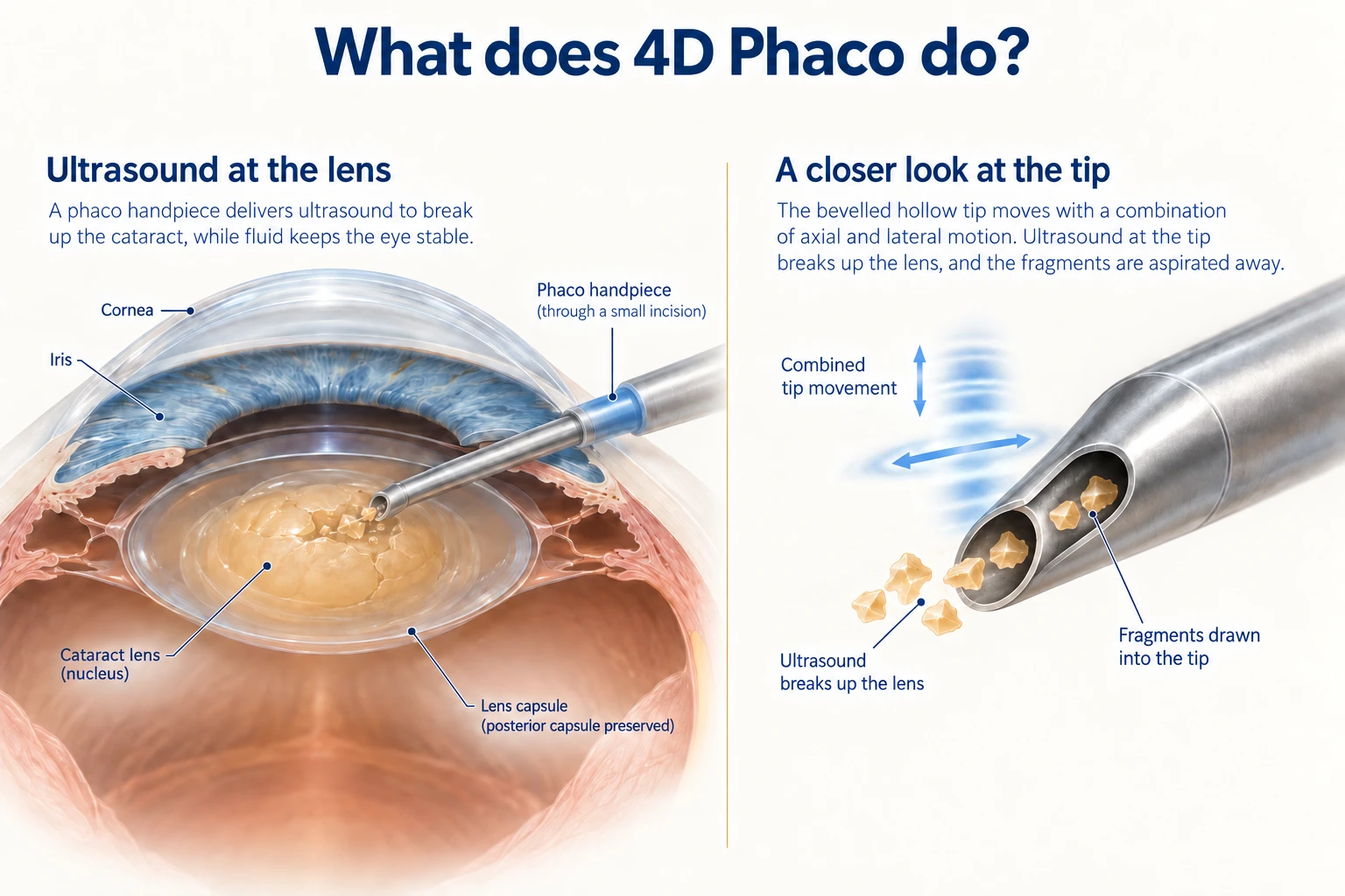
4D Phaco is Alcon's ultrasound technology for breaking up the cataract. It is not a four-dimensional scan. “Phaco” is short for phacoemulsification: fragmenting the cloudy natural lens with an ultrasound-driven tip and drawing the pieces out.
The tip moves by tiny amounts at very high speed. In 4D Phaco, axial and lateral movement are combined with modulation of the tip's stroke. In everyday terms, the way the tip moves is designed to help it work through lens material efficiently. Fluid around the tip supports the eye; suction carries fragments through its hollow centre.
A useful distinction is between breaking material up and bringing it to the tip. Ultrasound does the first job; fluid flow and suction help with the second. They need to work together. More ultrasound power is not automatically better.
The aim of more efficient removal is to accomplish the necessary work with less unnecessary ultrasound exposure. That is a sensible engineering goal. Whether it produces a meaningful difference in corneal recovery, complications or final vision requires clinical comparison, discussed below.
This ultrasound function is also different from femtosecond laser-assisted cataract surgery, which can assist with selected earlier steps of a cataract operation. The two terms are not interchangeable.
Why are pressure, vacuum and flow different?
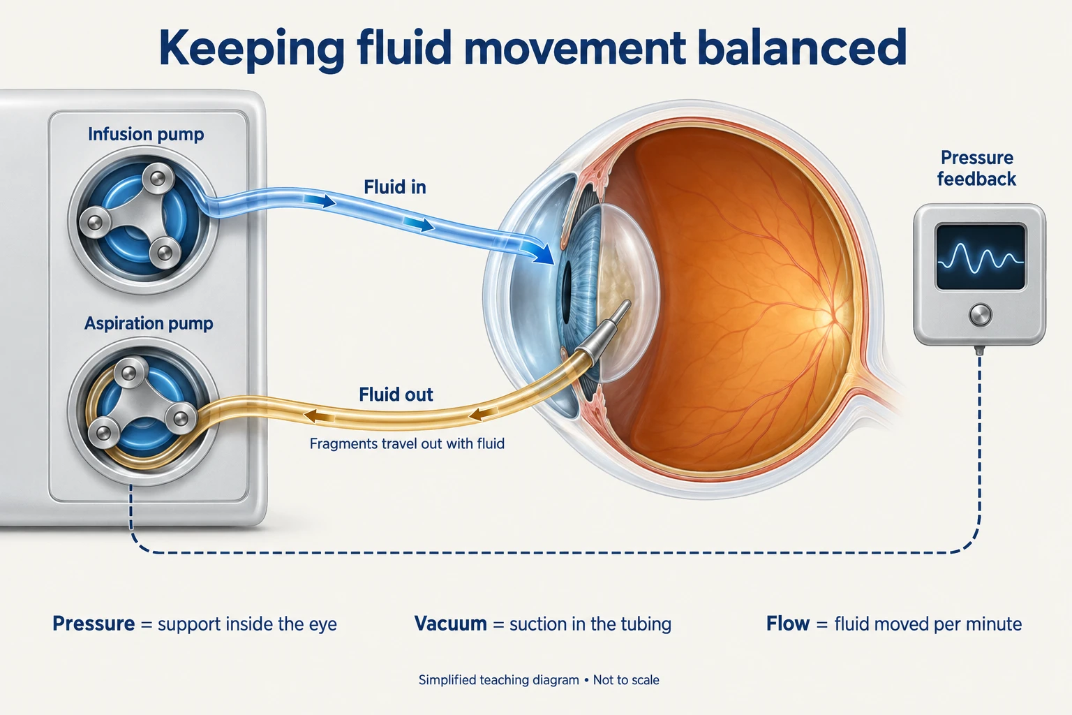
The eye needs to retain its shape while material is removed. Too much fluid leaving relative to fluid entering can reduce the working space; excessive pressure is also undesirable. Fluidics is the name for controlling this moving fluid.
Three measurements describe different parts of the job:
- Eye pressure, or IOP: the pressure supporting the eye from within.
- Vacuum: the suction generated in the aspiration system to hold and remove material.
- Flow: the volume of fluid moving in a given time, usually shown as mL/min or cc/min.
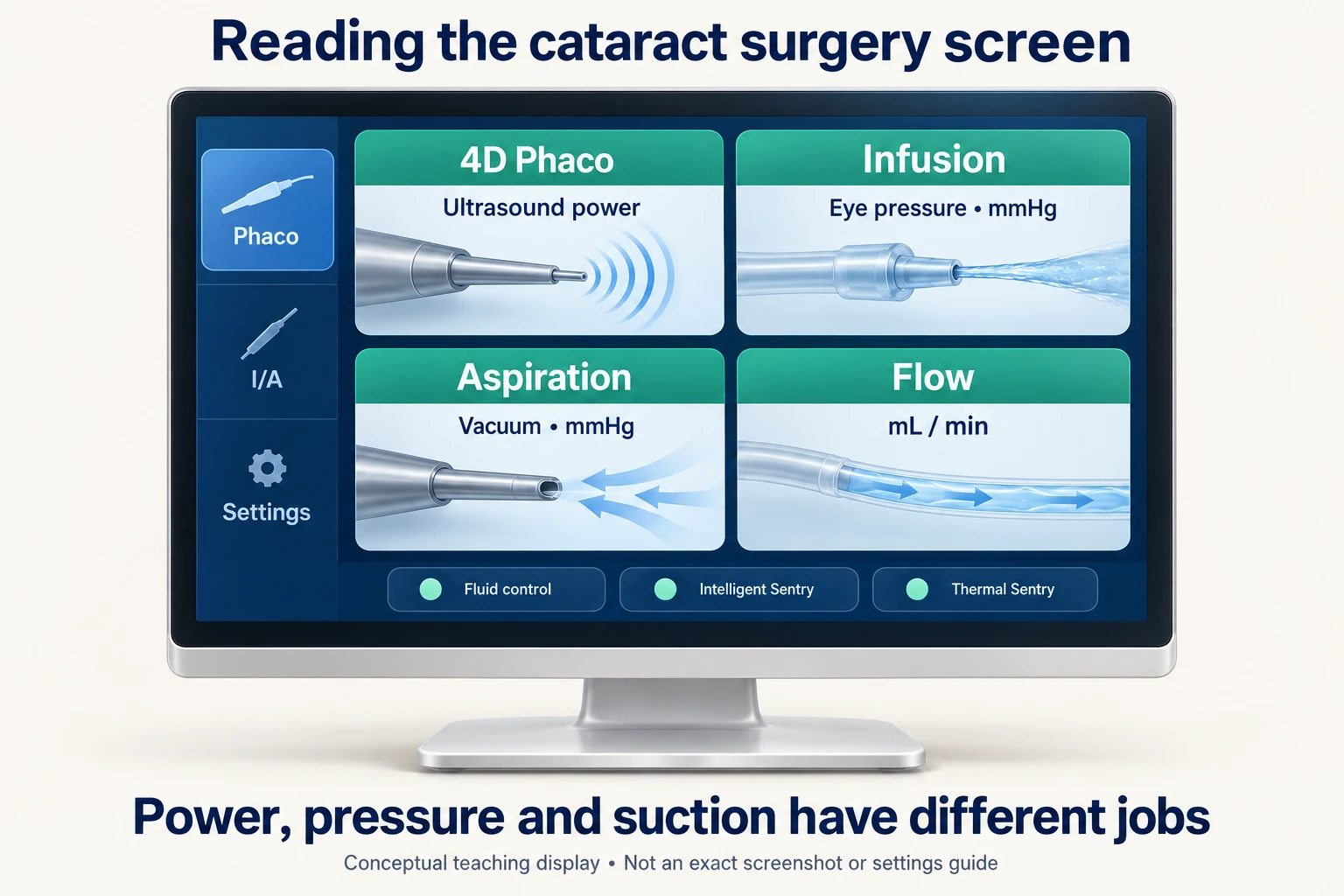
Both pressure and vacuum may be displayed in mmHg, but they are not the same measurement. A high vacuum value on the screen does not mean the eye itself is being held at that pressure.
UNITY's dual-pump arrangement controls fluid entering and leaving separately. Feedback allows the system to respond as resistance, leakage or demand changes. The purpose is to keep useful operating space while the surgeon removes material.
Intelligent IOP concerns pressure control. Intelligent Aspiration concerns how suction and flow respond to the task and foot-pedal input. Rapid suction can help engage material, while controlled flow matters when working near delicate structures. The settings still need to suit the operation and the tissue involved; there is no single ideal number for every patient.
What does Intelligent Sentry do?
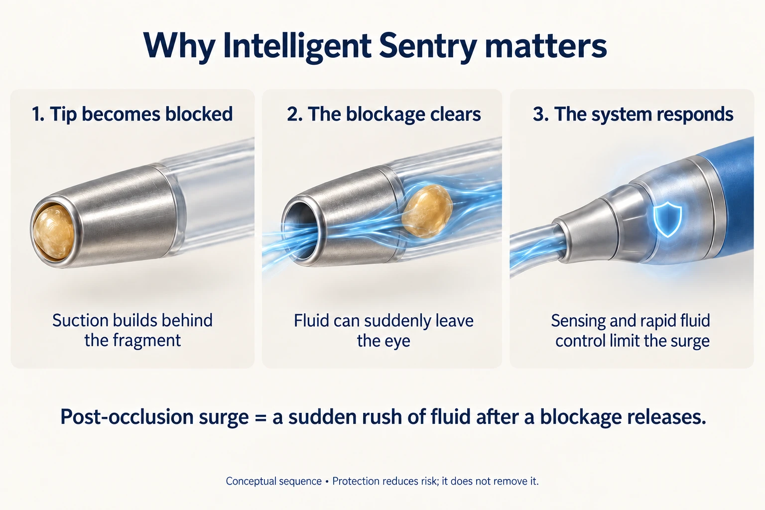
Intelligent Sentry helps control the sudden fluid movement that can follow a blocked phaco tip clearing.
A piece of cataract can temporarily seal the tip's opening. Suction builds behind it. When that piece breaks up or moves through, fluid can rush into the tubing. This is called post-occlusion surge: “occlusion” means blockage, and “surge” means a sudden rush.
The practical concern is a momentary loss of fluid and operating space inside the eye. Intelligent Sentry uses sensing and a rapid response involving vent valves to mitigate that event. A vent valve helps release suction when required.
This is a fast response to a particular event, alongside the broader fluid-control system. The surgeon must still watch the chamber, position the instruments and adjust the technique when necessary.
How is Thermal Sentry different?
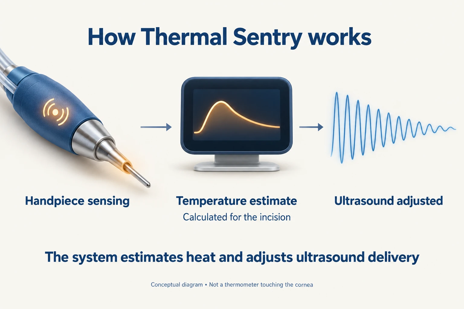
Thermal Sentry addresses heat during ultrasound use. It has a different job from Intelligent Sentry's surge response.
Ultrasound delivery and movement at the instrument–incision interface can generate heat. Irrigation and appropriate technique help control it. A significant thermal injury to a cataract incision is uncommon but can affect the wound.
Thermal Sentry uses information from handpiece sensing and an algorithm to estimate incision temperature, then regulate ultrasound delivery. Think of it as an additional feedback mechanism for heat. It is not a thermometer sitting against the cornea, and it does not replace adequate irrigation or careful instrument use.
What do the cataract surgery screens show?
The display separates ultrasound, fluid support and aspiration controls. The team can see the active mode and relevant settings, while the surgeon varies instrument action with the foot pedal. During irrigation and aspiration, often abbreviated I/A, fluid and suction remove remaining lens material without the same ultrasound fragmentation step.
The screen is part of the team's situational awareness, alongside direct observation of the eye. A reassuring number does not take precedence over what the surgeon sees. Guided setup and component checks help the theatre team prepare the system, but pre-operative checks and staff familiarity remain necessary.
What changes when the machine is used for retinal surgery?
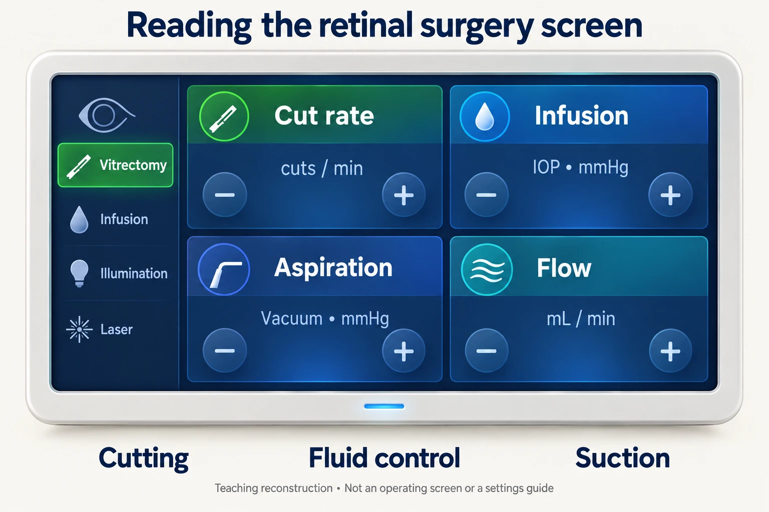
In vitrectomy, a fine cutting probe removes vitreous gel from inside the eye. The goal may be to gain access to a retinal problem or relieve traction. The required operation depends on the diagnosis; removing gel is only one part of it.
The compatible HyperVit dual-blade probe can operate at up to 30,000 cuts per minute. Its small internal cutting mechanism divides material as it is drawn into the probe. Cutting the gel into smaller portions helps the surgeon manage its removal. The probe does not spin around freely inside the eye, and the number of cuts per minute does not tell you how long the whole operation will take.
Dynamic Stiffener addresses another practical issue: very fine instruments can flex. The 27+ DS instruments add support to help the surgeon control these small tools. In surgical instrument sizing, a higher gauge number generally means a narrower instrument. Smaller does not automatically mean better for every task.
TetraSpot is a compatible retinal laser probe that can deliver selected patterns of one, two or four spots. Multiple spots can reduce repetitive positioning when treating a suitable retinal area. This is laser treatment inside the eye, not laser cataract surgery. The surgeon still decides where treatment is needed, which pattern to use and how to avoid structures that should not be treated.
The vitrectomy guide explains why retinal surgery is performed and what recovery may involve. These instruments support the operation; they do not determine whether an individual retinal condition can recover.
What does Light Sentry do?
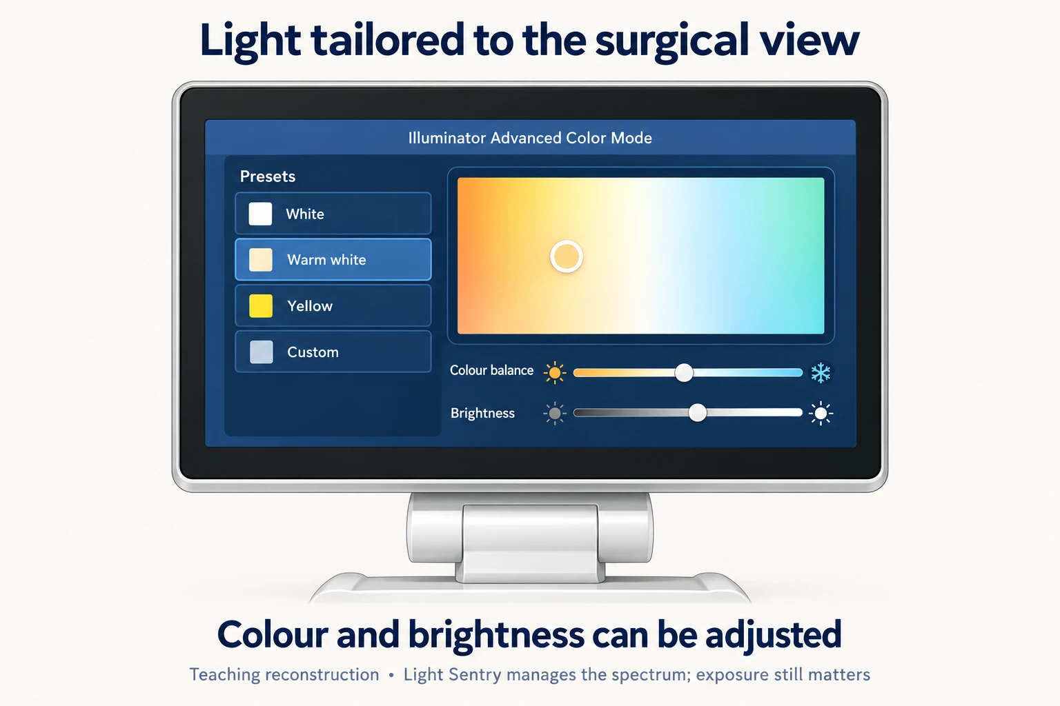
Light Sentry relates to the illumination used to see inside the eye during retinal surgery. A fine light probe illuminates the surgical field. UNITY's LED illumination allows adjustment of colour and brightness to suit the view.
Light Sentry manages the spectrum—the mixture of wavelengths in that light—to address potentially harmful exposure. This matters because the retina is light-sensitive. Exposure still depends on brightness, duration, probe position and other factors. The feature does not remove the need to manage light carefully.
The three names can therefore be kept distinct: Intelligent Sentry manages surge, Thermal Sentry manages heat, and Light Sentry manages the light spectrum.
What benefits have actually been studied?
The evidence supports technical and workflow benefits more clearly than a promise of better vision for every patient.
A 2026 pressure-control paper included laboratory testing and a 68-patient cataract series. Surgeons reported stable chambers in that series, but there was no clinical comparison group. It cannot establish fewer complications than another machine.
A separate 2026 experiment compared ultrasound methods using portions of one very dense cataract nucleus outside the body. Its efficiency findings are interesting, but a single specimen cannot predict patient outcomes.
A retinal study published in September 2026 analysed 179 operations. Total console-associated workflow averaged 27.3 minutes with UNITY and 32.4 minutes with CONSTELLATION. That measure included setup, the procedure and teardown. There was no statistically significant difference in the overall intraoperative duration. The comparison was observational, Alcon sponsored, and excluded combined cataract and retinal operations.
These are useful pieces of evidence, with different questions and limitations. They do not justify converting a bench efficiency percentage into a percentage reduction in surgical risk. Some early research and many technical claims also involve the manufacturer, which is relevant when judging how independent the evidence is.
What should patients and optometrists take from this?
UNITY VCS offers several ways to help a surgical team control how an operation is performed. The potential value is in efficient tissue removal, responsive fluid management and useful retinal instruments. The right use of those capabilities still depends on the surgeon, the theatre team and the individual eye.
For a patient, useful questions are which procedure is recommended, what is limiting vision, what risks apply to that eye and what recovery is realistic. For an optometrist, the important distinction is between a platform's capabilities and evidence that a particular patient group has better outcomes with it.
Cataract density, corneal health, pupil size, capsule support, retinal disease and previous surgery can all affect planning. The machine also does not determine lens implant choice. Where both cataract and retinal surgery are needed, the decision about combined or separate surgery requires its own assessment.
A technical note for optometrists
When reading a device comparison, check whether it measured steady-flow pressure, transient surge, an ultrasound energy index, endothelial cells, vision or theatre time. These endpoints are not interchangeable. Ask whether the comparator used similar settings and whether the study was a bench experiment, a single-arm series or a randomised clinical trial. This helps explain new technology to patients without overstating what has been established.
Sources and further reading
- Alcon: UNITY VCS and CS product information . Manufacturer descriptions and specifications.
- Alcon: UNITY launch information, April 2025 . Manufacturer technical overview.
- Alcon: technical account from Duke Eye Center . Manufacturer-hosted account; not independent comparative evidence.
- Berdahl and colleagues: pressure control and anterior chamber stability . Clinical Ophthalmology, 2026.
- Giglio and Tognetto: a single-nucleus comparison of ultrasound methods . Frontiers in Ophthalmology, 2026.
- Busquets and colleagues: comparative retinal surgical workflow . Ophthalmology and Therapy, 17 September 2026.
Information and sources checked on 21 September 2026. Illustrations are educational; they do not show a patient's eye or prescribe surgical settings.