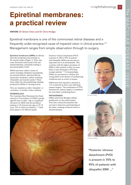
The macula is the small central area of the retina responsible for detailed vision. An epiretinal membrane is a thin sheet of scar-like tissue that grows on its surface.
01 Macula · surface detail
The macula and its surfaceThe membrane lies on the surface of the macula.
Illustrative schematic · not to scale
02
How a membrane changes the surface
As the membrane contracts it puckers the retina beneath, like cling film tightening over a surface, which blurs and distorts central vision. It is sometimes called macular pucker or cellophane maculopathy. Most membranes are related to the normal ageing separation of the vitreous gel from the retina; some follow a retinal tear, retinal surgery, inflammation or a blood vessel problem in the eye.
02 Macula · surface detail
How a membrane changes the surfaceAs a membrane contracts, it puckers the retina beneath.
Illustrative schematic · not to scale
03
Measuring changes over time
Many membranes are mild, change little and are simply watched. Others thicken and contract over months or years. The membrane is diagnosed and measured with an OCT scan, and it can be removed surgically when it is affecting vision enough to make the trade-offs worthwhile.
03 Macula · surface detail
Measuring changes over timeAn OCT scan maps the layers of the macula in cross-section.
Illustrative schematic · not to scale
01 Macula · surface detail
The macula and its surfaceThe membrane lies on the surface of the macula.
Illustrative schematic · not to scale
02 Macula · surface detail
How a membrane changes the surfaceAs a membrane contracts, it puckers the retina beneath.
Illustrative schematic · not to scale
03 Macula · surface detail
Measuring changes over timeAn OCT scan maps the layers of the macula in cross-section.
Illustrative schematic · not to scale
In brief
Affects central vision only; side vision is unaffected
Often progresses slowly and can be watched
Diagnosed and measured with an OCT scan
Vitrectomy with membrane peel is the operation
Surgery is for vision that matters to you, not for the scan alone
How vision changes
Symptoms of an epiretinal membrane.
⇔
ClearWith a membrane
Straight lines bend
Tiles, door frames, window bars and lines of print look wavy or kinked, and objects can seem the wrong size or shape in the affected eye.
⇔
ClearWith a membrane
Gradual central blur
The centre of vision softens over months, so small print and faces lose their sharpness. Glasses do not remove distortion caused by the membrane, although they may help other focusing problems.
Usually one eye
Noticed by chance
Most people notice the problem in one eye, often when the other is covered. The good eye tends to compensate, which is why the change can go unnoticed for a while.
Slide each picture to compare typical vision with the change described. Illustrations are indicative, not a diagnostic test or a prediction of vision after treatment.
At the consultation
The OCT scan tells the story.
An OCT scan (optical coherence tomography) maps the layers of the macula in cross-section, showing the membrane and the wrinkling beneath it.
After a vision test and dilating drops, the macula is examined and scanned. The scan is repeated over time to measure change, which guides the decision about whether and when to operate. Because the lens sits in front of the retina, any cataract is assessed at the same time; the two often need to be planned together.
Dr Chen explains what the scan shows and whether watching, or surgery, is the sensible next step for that eye.
When surgery is considered
When distortion or blur is affecting reading, driving or daily life, rather than simply because a membrane is present
When the scan shows the membrane thickening or the retina swelling beneath it
When a cataract is also due, so both can be planned as one problem
Suitability is individual and decided at the consultation.
The operation
Vitrectomy and membrane peel, in plain terms.
The operation is called vitrectomy with membrane peel. It is delicate, precise work under the microscope, usually as day surgery.
Through three tiny openings in the white of the eye, the vitreous gel is removed (a vitrectomy). The membrane is then lifted off the surface of the macula with fine forceps, often after a dye is used to make it visible. The openings are usually small enough to seal without stitches. A gas bubble is sometimes placed in the eye at the end; for a membrane alone it is often not needed.
Surgery is under local anaesthetic with sedation, or general anaesthetic when that suits the person. Dr Chen is a vitreoretinal surgeon, a surgeon of the retina and the vitreous gel, and performs this operation regularly.
Contains surgical footage. An existing educational example from Dr Chen’s public channel.
Risks and limitations, in plain language
Complications are uncommon but include infection, bleeding, retinal tear or detachment, raised pressure and, rarely, loss of vision. A cataract commonly develops or advances after vitrectomy in eyes that still have their natural lens, so cataract surgery is often planned alongside. Distortion usually improves but may not disappear, and vision continues to settle for months. All of this is discussed with you before surgery.
General information, not a guarantee of outcome.
After surgery
What recovery looks like.
Improvement is gradual. Vision after a membrane peel continues to settle over months rather than days.
Drops and care. Drops for several weeks; avoid rubbing the eye, swimming and dusty environments for the period advised.
If gas is used. Vision is blurred while the bubble is present and clears as it absorbs over weeks. No flying or travel to altitude until it has gone, and tell any anaesthetist about the bubble.
Everyday activities. Reading, screens and gentle walking are fine once comfortable; heavy lifting and strenuous exercise wait for the period advised.
Distortion. Often improves gradually; some can remain, and what is realistic for your eye is discussed beforehand.
Reviews. Arranged through Vision Eye Institute, with a repeat OCT scan to confirm the macula has settled.
Between visits
Report any sudden loss of vision, increasing pain or redness, or a new shadow in the vision rather than waiting for the next appointment.
Read an article about epiretinal membranes written by Dr Simon Chen with Dr Chris Hodge, covering assessment and treatment in more detail.
The original published article for eye-care professionals.
Questions to ask
Useful questions for the consultation.
Is the membrane affecting my vision now, or is it something to watch?
What does my OCT scan show, and how will we measure change?
If surgery is suggested: what improvement is realistic for my eye, and what may not improve?
Will I need a gas bubble, and will I have to keep a head position?
Should the cataract be dealt with at the same time?
What are the risks for my eye in particular?
How long before I can drive, work and read comfortably?
Bring these, and anything else on your mind. Write the answers down, or bring someone who can.
Common questions
Straight answers, without replacing a consultation.
Do all membranes need surgery?
No. Many are mild and stable, and are simply watched with OCT scans. Surgery is considered when the membrane is affecting vision in a way that matters to you.
Will my distortion go completely?
Distortion often improves after surgery, but some can remain, particularly when a membrane has been present for a long time. What is realistic for your eye is discussed candidly before surgery.
Will I need cataract surgery as well?
Often, yes. Vitrectomy accelerates cataract in eyes that still have their natural lens. Because Dr Chen performs both, the cataract can be planned at the same operation or in sequence, whichever suits the eye.
Can a membrane come back?
Regrowth after a peel is uncommon. The eye is reviewed with OCT after surgery to confirm the macula has settled.
Is a membrane the same as a macular hole?
No. A membrane sits on the surface of the macula; a macular hole is a small gap in the centre of it. They are related, sometimes occur together, and are both treated with vitrectomy, but the decisions differ. See the macular hole page.
Ready when you are.
Appointments and enquiries through Vision Eye Institute, Chatswood.